Position
Copyright 2009
Introduction
The heart lies between the lungs in the middle mediastinum and is enclosed in the pericardium. It is placed obliquely in the chest behind the body of the sternum and adjoining parts of the rib cartilages, and projects farther into the left than into the right half of the thoracic cavity. About one-third of it is situated on the right and two-thirds on the left of the median plane. Boundaries of the heart are clinically important as an aid in diagnosing heart disorders.
Position of the Heart |
|
The heart lies within the chest cavity slightly to left of midline, in the lower 2/3, and rests on the diaphragm. 45772 Courtesy Ashley Davidoff MD Davidoff art copyright 2009 |
The heart does not lie upright with atrial chambers superiorly and ventricular chambers inferiorly. Rather it is tipped with the atria rightward and backward, and ventricles leftward and forward. In addition, the right sided chambers are more anterior and superior than left sided chambers. It is of extreme importance for you to spend some time on this section trying to visualize in your mind’s eye how the heart lies, since once the template is captured, you can turn the heart in any which way and you will be able to find your way around it.

Orientation and Parts of the Septa of the Heart
|
|
The heart chambers do not lie in a vertical orientation. The atria are rightward, superior and posterior to the ventricles. The right sided structures are anterior and superior to the left sided structures. 01667b14 Davidoff drawing Davidoff MD |
A poem entitled The Heart of the Fair Young Maiden explains the positioning of the atria and ventricles
The relevance of the anatomy is introduced in the evaluation of the chest X-ray
 
The Lateral Chest Examination of the Heart Relative Position |
|
The right ventricle is anterior to the left, and is also superior. The right atrium (blue ring overlay) is slightly superior and anterior to the LA . ie Blue right sided structures are superior and more anterior than red structures . The atria are superior and posterior to the ventricles as shown in the diagram. Courtesy Ashley Davidoff all rights reserved copyright 2009 15416 C 02 W.8s, 15416 C 02 W.82s |

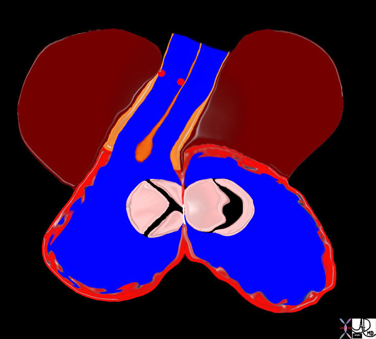
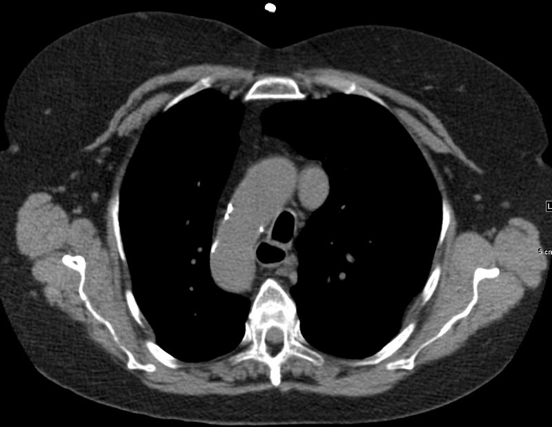
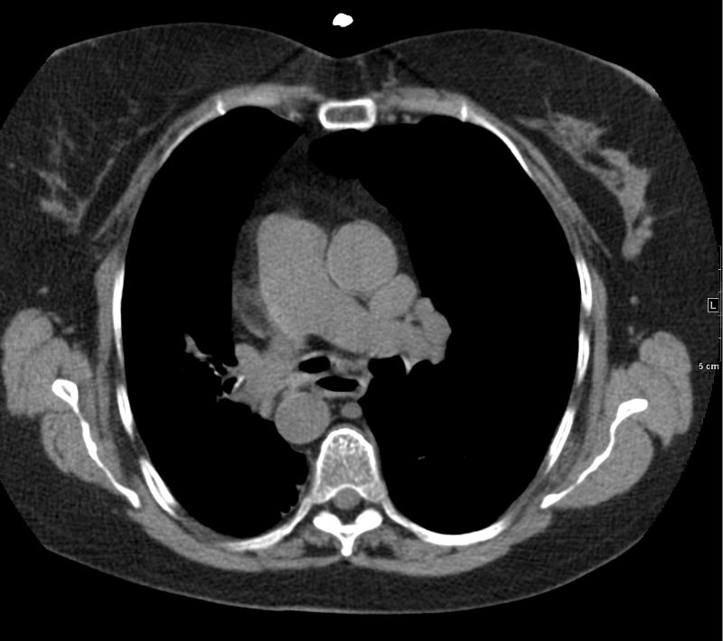
Position of the Chambers in Relation to Each Other in the Axial Plane
|
|
The cross sectional image from a CT scan through all 4 chambers reveals the relative position of the chambers and A-V valves. In general “blue” chambers are both anterior to and superior to the “red” chambers so that both the right atrium (RA) and right ventricle (RV) are anterior to the LA and LV. The tricuspid valve (blue valve) follows the rule and is slightly anterior to the mitral valve as well. Courtesy Ashley Davidoff Copyright 2009 all rights reserved The atria are posterior to the ventricles. 27531cd01.8s |
Embryologic Considerations.
The position of the heart in the chest, and the position of the chambers are defined in the embryological development of the heart.
There are 4 major events in embryology of the heart.
Situs development
Looping
Septation
Growth and regression
Positioning of the apex is a minor independant event separate from situs and and looping.
Of these events situs, looping, and apex positioning play a major role in the positioning of the heart and its components.
Innate in the genetic instruction is positioning of the atria, lungs tracheobronchial tree and some of the abdominal organs. Normal situs instruction would result in the positioning of the right atrium on the right, left atrium on the left, right lung, liver, pancreatic head, and gallbladder on the right, and left lung spleen, and pancreatic tail on the left.
Looping of the heart is also under genetic instruction, and normal looping would bring the right ventricle to the right and left ventricle to the left. This usually places the aorta to the right of the pulmonary artery.
Positioning of the apex is independant event and is usally the result of the final positioning of the left ventricle.
|
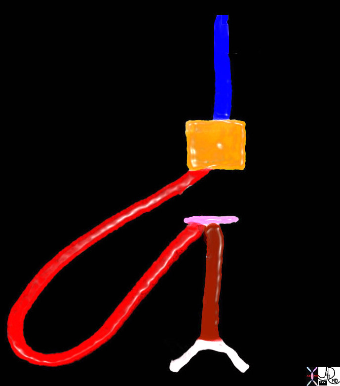
Rightward Looping of the Heart |
| The right ventricle is the first ventricle to develop and it loops to the right and anteriorly, leaving the developing atria posteriorly. This diagram shows the right ventricle (bright red) taking a a loop to the right, and is called a d loop for dextro. This event is independant of situs development which positions the atria.
01488b02 Davidoff Art Davidoff MD Davidoff art drawing Davidoff MD |
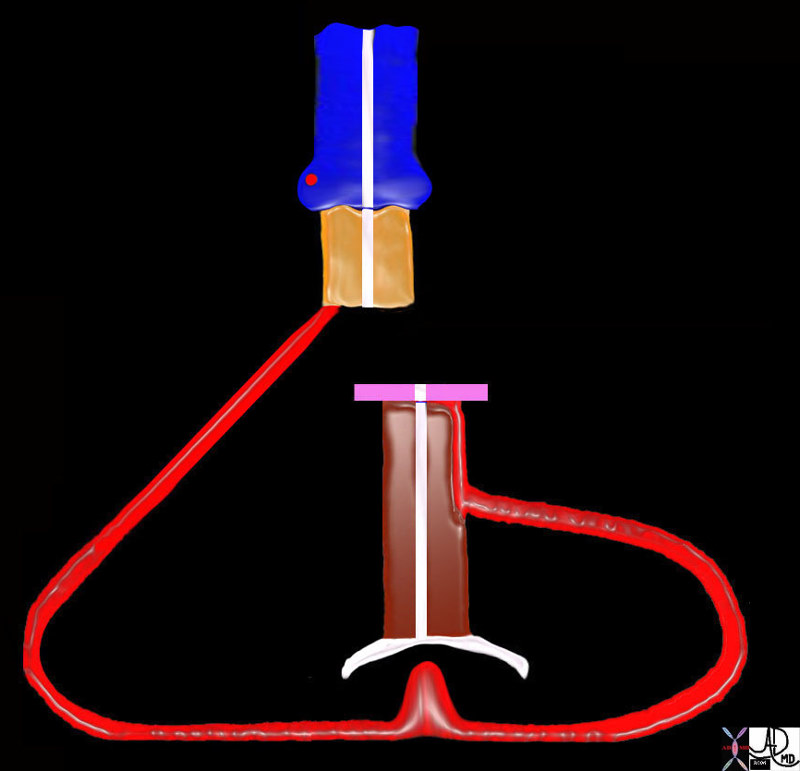
Development of the Left Ventricle |
| The “D” loop defines where the right ventricle is going to be positioned, and the left ventricle develops as a daughter ventricle off the right. In the normal development the d loop results in a normal positioning of the left ventricle, posterior to the right and anterior to the developing atria.
01477e03 Davidoff art drawing Davidoff MD |
|
Apex |
| Septation divides atria, ventricles, great vessels, and A-V canal. As an independent event the apex is normally positioned by the LV toward the left side, creating levocardia.
01492b14 Davidoff art Davidoff MD copyright 2009 |
Levocardia
Levocardia is the normal positioning of the heart dominantly in the left chest as a result of the normal placement of the apex by the left ventricle
Levocardia |
| Normally positioned apex – as a result of a D loop and settling of the apex into the left chest, as its final position.
45756 Courtesy Ashley Davidoff MD Copyright 200945756 45757 |
Dextrocardia
Dextrocardia is defined as a malposition of the heart that can be congenital or acquired resulting in the apex of the heart being in the right chest. There are two congenital forms; One is associated with situs solitus, and the second associated with situs inversus. The form that is associated with situs solitus is thought to be due to an embryonic arrest resulting in the the right ventricle (rather than the left) forming the apex. In this instance the right ventricle is still on the right side and the left ventricle is on the left side. This form of dextrocardia is associated with a higher incidence of congenital abnormalities. The second form of the congenital version is associated with situs inversus. In this situation the incidence of associated congenital abnormalities is lower.
Acquired dextrocardia may be due to either loss of volume in the right chest or a push due to mass effect from the left side.

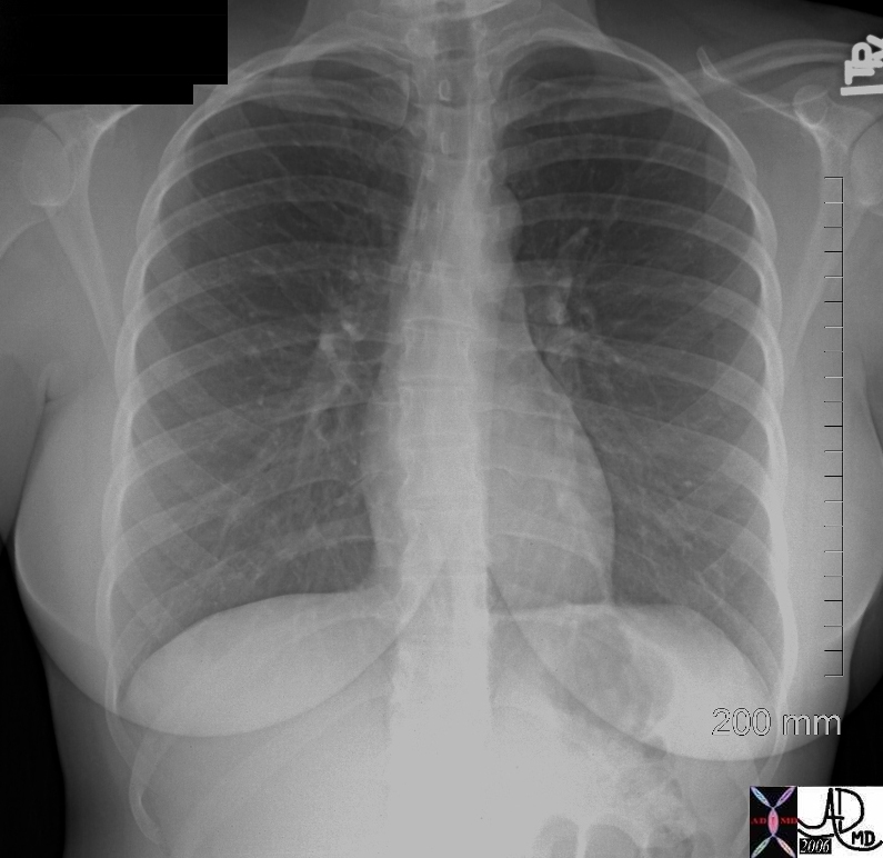
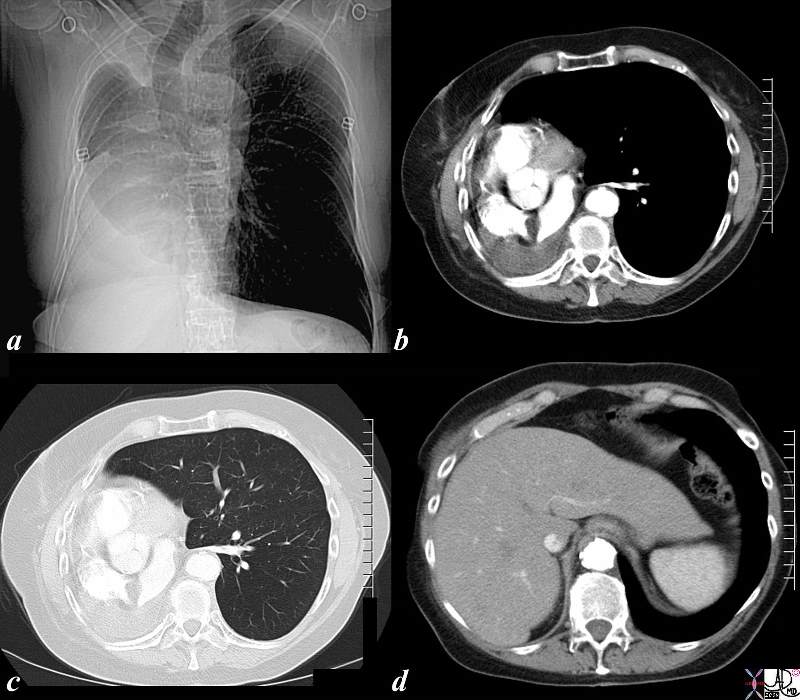
Dextrocardia |
|
The PA chest X-ray shows the apex pointing rightward and most of the heart is in the right chest. Note also that the stomach bubble is in normal position, and so situs solitus is present. This is a case of isolated dextrocardia, caused by an arrest of development, not associated with situs inversus, but subject to a higher incidence of other congenital anomalies. (Images courtesy of Ashley Davidoff M.D.) |
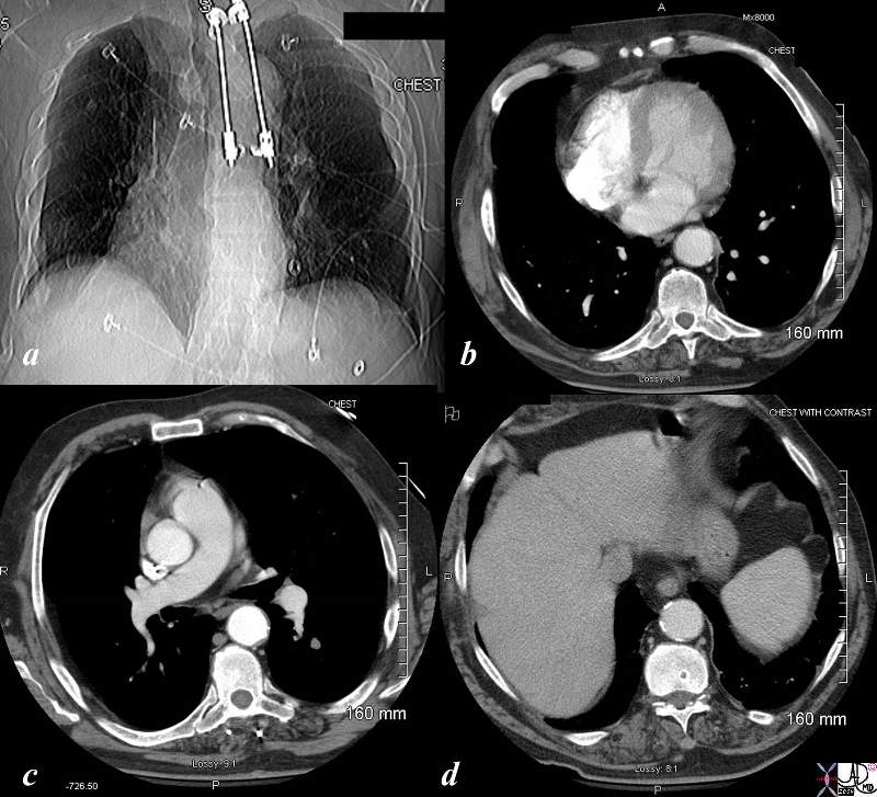
Dextrocardia with Situs Solitus |
| The CT is from an elderly patient with dextrocardia and situs solitus. The scout film before the CT shows dextrocardia and Harrington rods are noted in the thoracic spine. Image b, shows the apex of the heart pointing to the right, but less impressive than the plain film. Image c through the great vessels show normal relationship, and through the abdomen, situs solitus is confirmed.
Courtesy Ashley Davidoff MD copyright 2009 45806c01.8s |
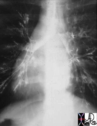
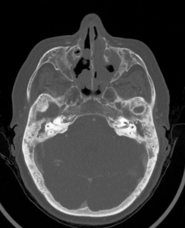
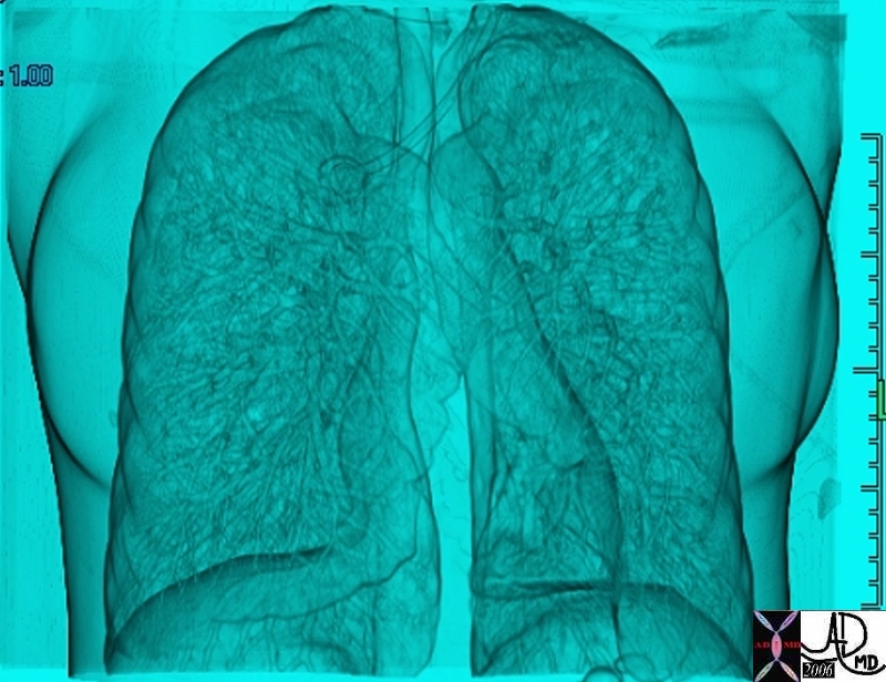
Dextrocardia and Situs Inversus |
| The CXR and bronchogram is from a 30year old male who presents with a long standing history of a productive cough, sinus headaches, and infertility.
The bronchogram shows inversion of the bronchi with the long thin bronchus characteristic of the left bronchus being right sided and short “fat ” bronchus characteristic of the right being left sided. Situs inversus and dextrocardia exist. However the history of sinusitis and infertility make the diagnosis of Kartagener’s syndrome likely. 06248 Courtesy Ashley Davidoff Copyright 2009 all rights reserved |
Acquired Dextrocardia |
| The series of CT scans show an acquired dextrocardia. In image a, the heart is not seen at all – but since it is not in the left chest, it is presumed to be in the right. The mediastinum as seen by the trachea is shifted to the right and the right lung is shifted across the midlineinto the left chest. the patient is status post pneumonectomy. Image b shows the heart completely in the right chest, and image c, shows the shift of the left lung across the midline and complete absence of the right lung. Image d confirms situs solitus
Courtesy Ashley Davidoff copyright 2009 all rights reserved 86476c01.8s |

Spurious Dextrocardia |
| In this case the scout film manifests with what appears to be a case of dextrocardia. Image b shows a slight of the heart to the right but the appearance is dominated by by a large right atrium and left atrium. Image c confirms the leftward position of the apex and normal relationship of the left apex and right ventricle. Image d shows situs solitus with reflux of contrast into the dilated hepatic veins indicating tricuspid regurgitation.
Courtesy Ashley Davidoff MD copyright 2009 48098c02 |
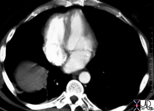
Mesocardia |
| This is a CT scan through the chest in which the apex of the heart points forward. The left ventricle (LV) is left sided and the right ventricle is right sided. There is situs solitus of the atria. This is a case of mesocardia ie neither right nor leftwad pointing apex.
Courtesy Ashley Davidoff MD. 19689 code heart mesocardia cardiac imaging radiology CTscan |
Situs Inversus
Situs inversus is a congenital disorder in which the organs of the chest and abdomen are inverted in position. Situs inversus is an abbreviated term for situs inversus viscerum.
The cause of the genetic aberration is usually an autosomal recessive but situs inversus is associated with entitites such as the asplenia syndrome, polysplenia syndrome, Kartageners syndrome (primary ciliary dyskinesia).
The result of the aberration is either a partial form (situs inversus partialis) or a total form (situs inversus totalis).
Structurally in situs inversus totalis the atria, tracheobronchial tree, and lungs are inverted, while in the abdomen the right sided organs including the liver gallbladder, head of the pancreas, and left sided structures such as the spleen, stomach, tail of the pancreas are inverted.
Situs inversus is not necessarily associated with dextrocardia. isolated dextrocardia without situs inversus is associatd with a much higher incidence of congenital heart defects.
Functionally situs inversus in the absence of other cogenital defects can be totally asymtomatic, but is usually fatal in asplenia syndrome. In this entity situs is usually ambiguus.
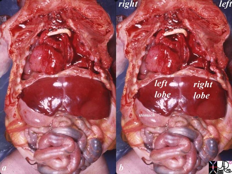
Situs Inversus Totalis, Dextrocardia and Severe Congenital Cardiac Anomalies |
|
The specimen is from a post mortem baby with severe congenital anomalies including situs inversus totalis, dextrocardia left sided liver right sided stomach severe congenital heart disease shunt possibly a Blalock Taussig large right ventricle grosspathology position malposition. Note that the right lobe of the liver is on the left, and the stomach is on the right. Courtesy Ashley DAvidoff MD copyright 2009 all rights reserved 10850c01.8s |

APEX POINTS TO RIGHT
SITUS INVERSUS OF BRONCHI,- HYPARTERIAL BRONCHUS ON RIGHT, EPARTERIAL BRONCHUS ON LEFT

TWO LEFT SIDED FISSURES AND DEXTROCARDIA

LIVER ON RIGHT , SPLEEN AND STOMACH ON LEFT