
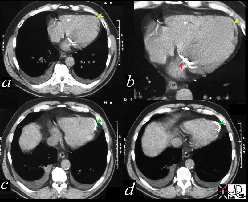
56 year old male with history of coronary artery disease. Axial CT through the heart shows apical curvilinear fat (yellow arrowheads, ( a and b) associated with apical myocardial dystrophic calcification (green arrowheads c and d) both indicating prior apical MI. In addition there mitral annular calcification (red arrowhead, b) and multifocal fatty deposits in the RV (white arrowheads, a and b) usually depicting age related degenerative changes,
Ashley Davidoff MD
Apical Hypertrophy and Mass Like Calcification –
heart0067-low res Ashley Davidoff MD

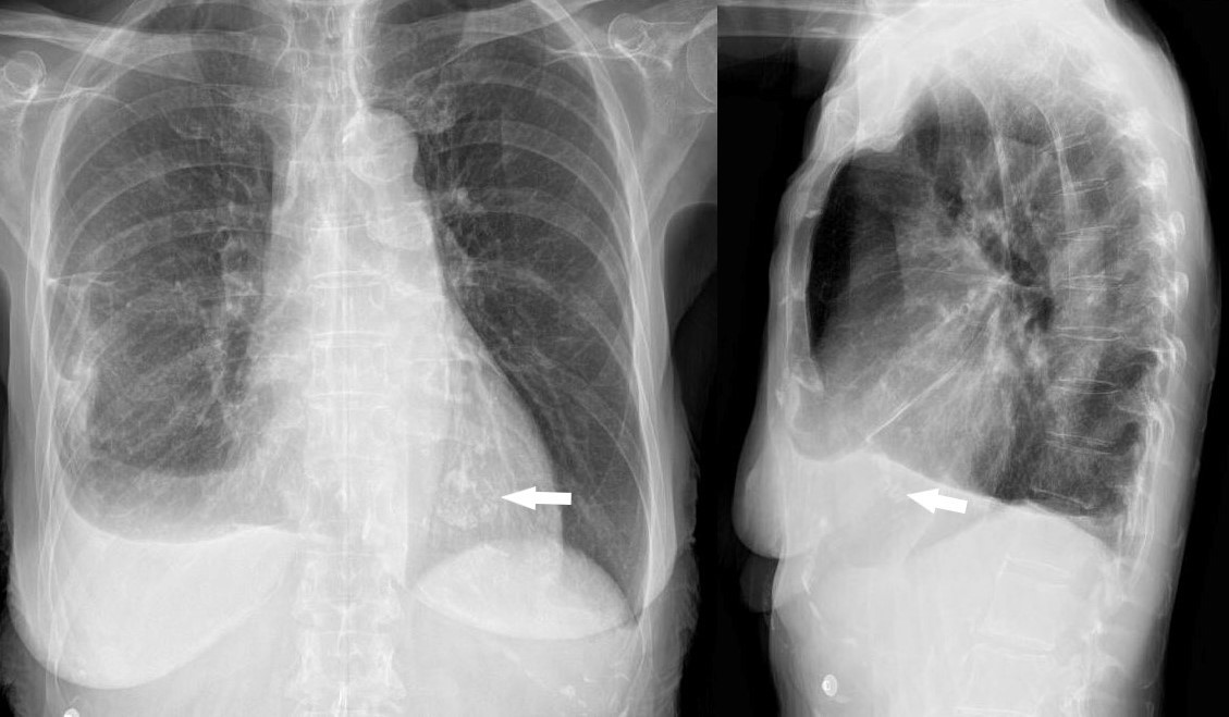
The patient is a 73 year old female with apical hypertrophic disease. She has a history of diastolic heart failure, pulmonary hypertension, CREST, esophageal stricture, COPD and chronic renal failure
heart0068b-low res
Ashley Davidoff MD
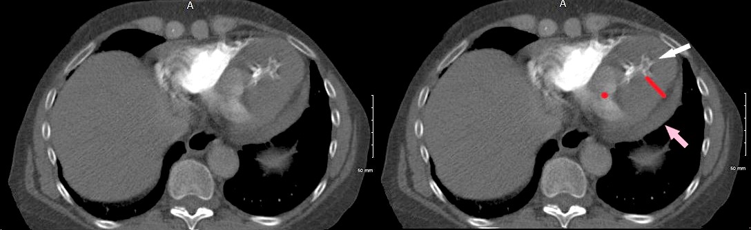
The patient is a 73-year-old female with apical hypertrophic disease. She has a history of diastolic heart failure, pulmonary hypertension, CREST, esophageal stricture, COPD and chronic renal failure
heart0069b02 Ashley Davidoff MD
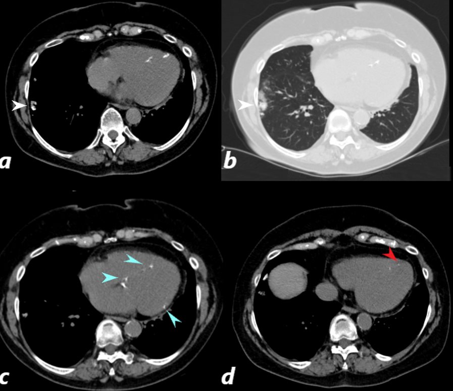
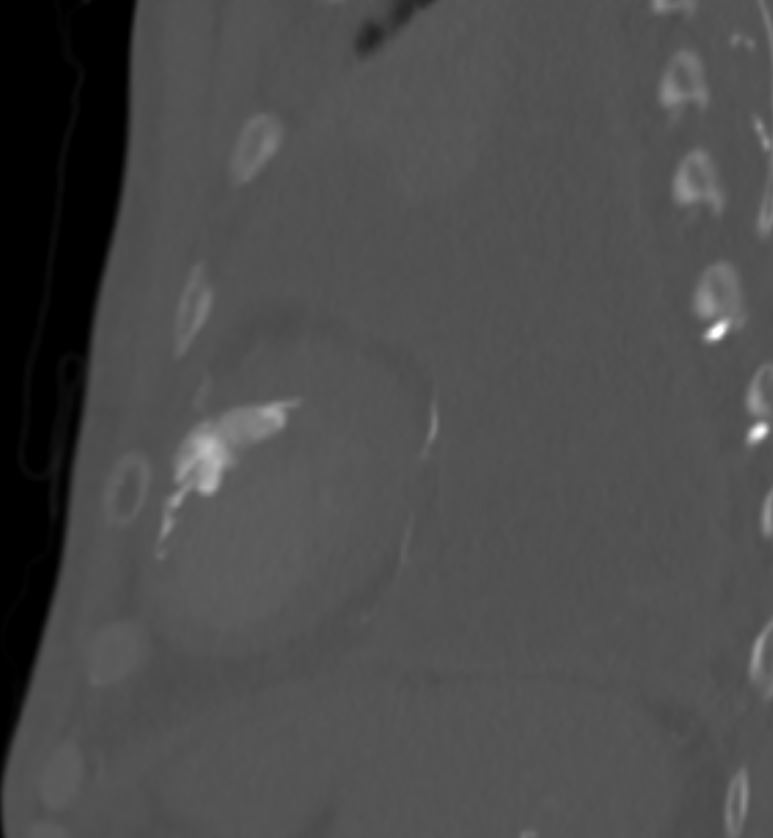
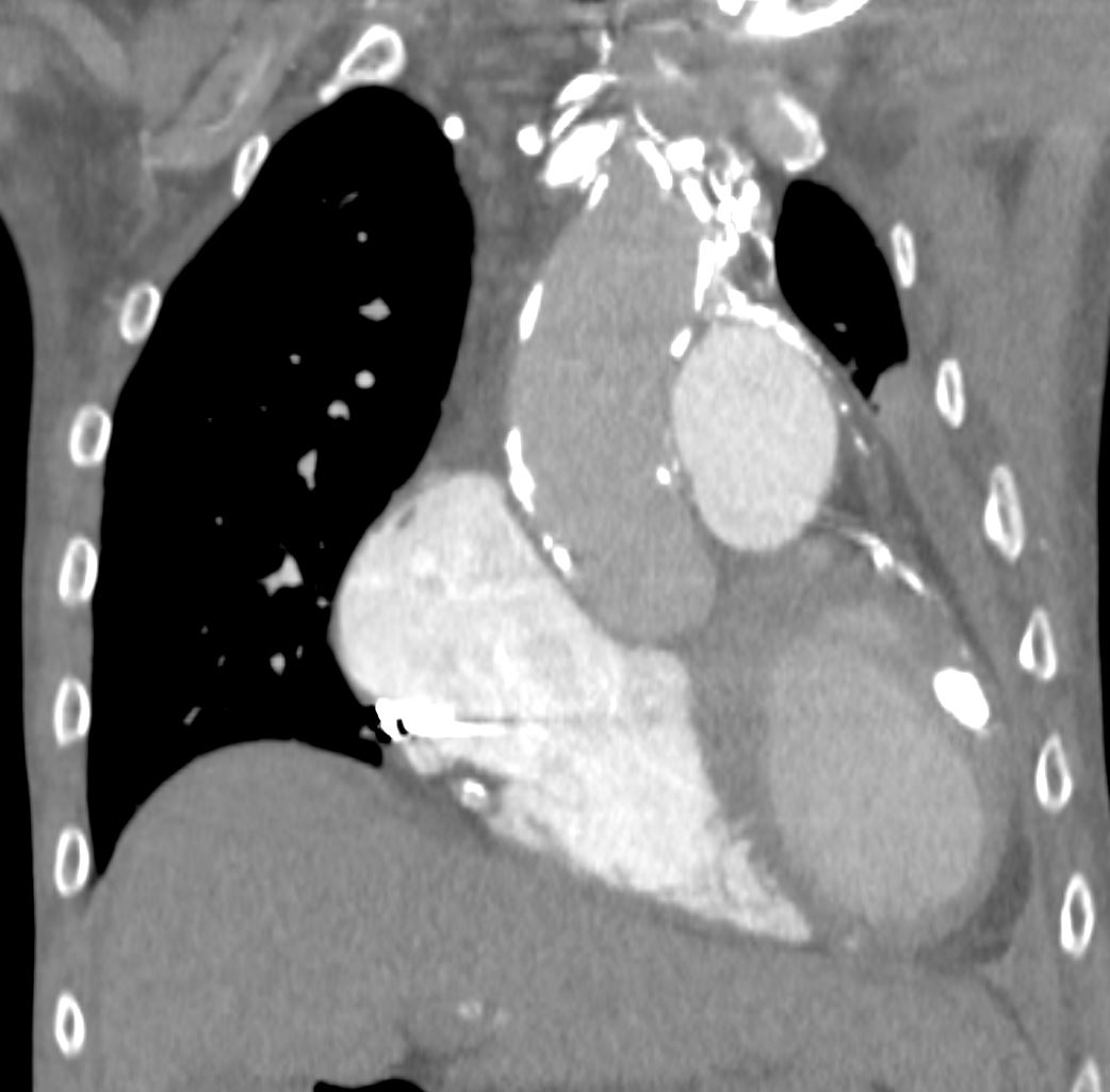
CT scan of a 67 year old female with anca vasculitis shows regions of dystrophic calcification in the lateral aspect of the right lower lobe (white arrow, a and b) )with focal nodular parenchymal consolidation, that likely reflects a site of prior small vessel infarct. Dystrophic calcification in the LV myocardium (blue arrows c) and a suggestion of fatty dysplasia in the left ventricular apex red arrow d) suggest changes from small vessel infarct.
Ashley Davidoff MD
Probable Dystrophic Calcification from Previous MI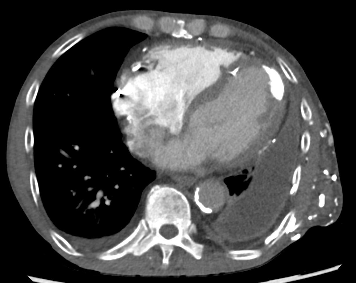

References
Khanna R kapoor A, Soni Neetu A Heart Set in Stone
Shim Ito et al Myocardial calcification with a latent risk of CHF in a patient with apical hypertrophic cardiomyopathy Internal medicine 54:1627-1631 2015