
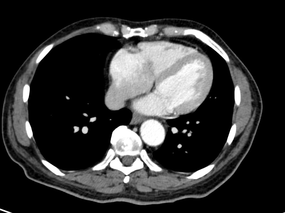
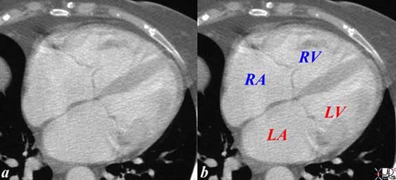
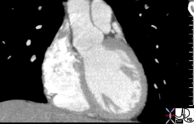
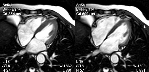
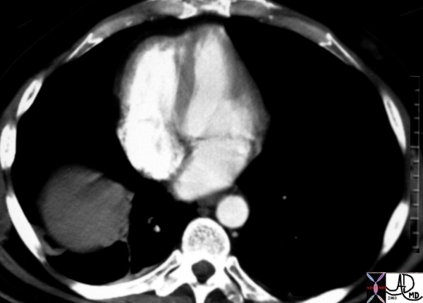
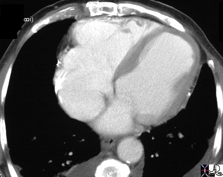
CT scan in the Axial plane of a healthy 24 year old male reveals a straightened RV side of the septum while there is a slight bulge on the LV side
Ashley Davidoff MD
The Common Vein Copyright 2019
Ganesh Athappan MD Ashley Davidoff MD
Definition:
The inter ventricular septum is a fibromuscular obliquely placed partition between the right and left ventricular cavities. Structurally it is composed of membranous and muscular parts and ,takes a gentle convex curvature toward the right ventricle. Functionally it serves to disconnect the right and left ventricular outflow tracts from one another .
Diseases of the ventricular septum are predominantly congenital with ventricular septal defect being the most common congenital heart disease .
Diagnosis of a VSD, is suspected in the presence of a thrill and a loud, harsh, or blowing holosystolic murmur over the lower left sternal border in the third or fourth intercostal space . The most common symptoms depending on the size of the defect are dyspnea, feeding difficulties, poor growth, profuse perspiration, recurrent pulmonary infections, and cardiac failure. The ECG shows equiphasic RS complexes in the mid precordial leads representing biventricular hypertrophy (Katz-Wachtel phenomenon ). The CXR may show RV enlargement and pulmonary plethora. Transthoracic echo by two-dimensional and Doppler color flow mapping identifies the type of defect in the ventricular septum in most cases. Cardiac catheterization may be used to determine the hemodynamic characteristics of the VSD.
Medical therapy for congestive heart failure or infective endocarditis, minimally invasive techniques and surgical options are available .
Structural Considerations
The infrastructure of the heart is built on cross, and the limbs of the cross reflect the septa of the heart. The upper septum called the atrial or interatrial septum divides the right and left atrium while the lower “vertical” septum divides the ventricles into right and left ventricles and is called the interventricular septum or simply the ventricular septum. The horizontal septum called the atrioventricular septum divides the atria from the ventricles. A third septum called the conal septum forms part of the interventricular septum as well as part of the septum at the base of the great vessels. The heart is tipped on its side with the apex pointing leftward and inferiorly while the right sided structures are tipped anteriorly and so the positional relationship of the structures are not orthogonal.
|
|

|
|
The atrial septum has three independent embryologic origins .
The upper portion arises from sinus venosus , the middle portion arises from the muscle itself (mesoderm), while the lower portion arises from the endocardial tissue.
|
|
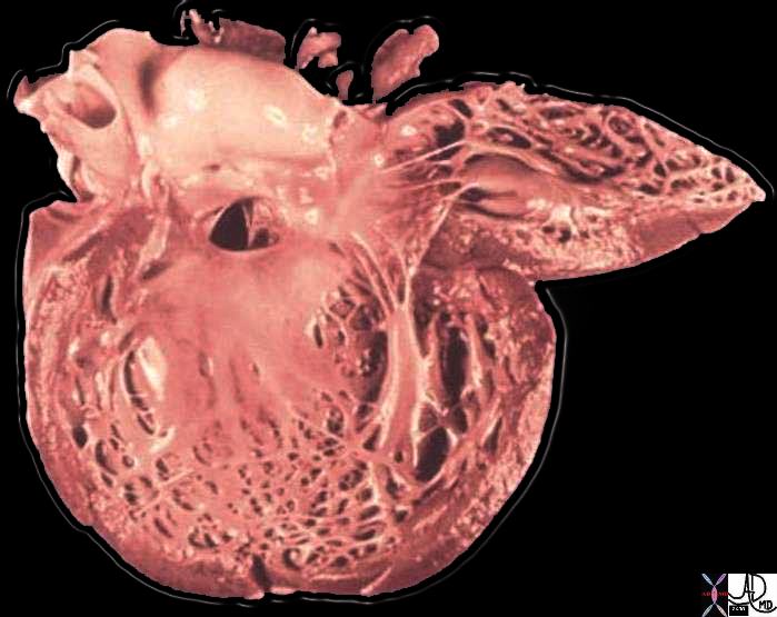
The strong ventricular septum, made up of a small membranous and large muscular portion is directed obliquely backwards and to the right, and is curved with the convexity towards the right ventricle. This septal geometry , subsequent to elevated LV pressures can be appreciated on imaging and is important as changes may signify either abnormalities of the septal myocardium or abnormal pressure differences between the LV and RV. Flattening of the interventricular septum may strongly correlate with RV overload.
|
|
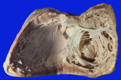
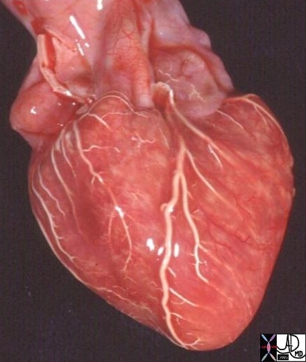 15009c cardiac heart coronary artery normal anatomy gross pathology LAD diagonal artery acute marginal artery conal artery arc of Vieussens Courtesy Ashley Davidoff MD 15009c
|
|
|
|
|
|
|
|
|
|
|
Imaging the Septum
CT scan in the Axial plane of a healthy 24 year old male reveals a straightened RV side of the septum while there is a slight bulge on the LV side
Ashley Davidoff MD
RV aspect :
Interventricular portion of the membranous septum: Part of the membranous septum that is overhung by the septal leaflet of the tricuspid valve.
Atrioventricular membranous septum: The part of the membranous septum that lies superior to the tricuspid valve and forms the floor of the right atrium.
Right ventricular Infundibulum : part of the right ventricle which lies between the pulmonary valve above and an imaginary line through the papillary muscle of the conus and upper edge of the membranous septum.
Crista supraventricularis : The crista supraventricularis if oversimplified can be considered synonymous with the infundibular (or conus) ventricular septum.
The appreciation of these terms is essential to understand the various classifications of ventricular septal defects which are discussed in the disease section of the cardiac module.
|
|
|
|
|
|

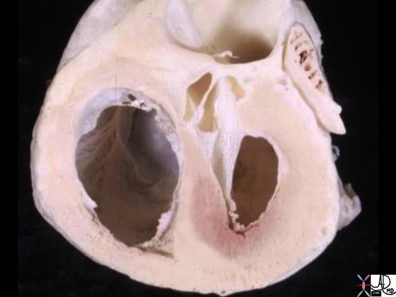
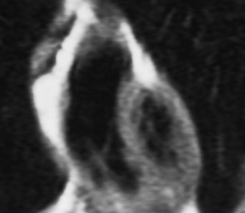
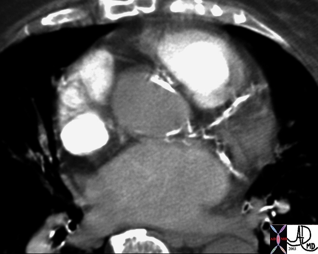
| Interventricular Septum As Seen In CT examination |
| 27531b01 right atrium heart cardiac RA tricuspid valve TV left atrium LA MV mitralvalve RV right ventricle anterolateral papillary muscle interventricular septum left ventricle LV CTscan Davidoff MD |
| Gentle Curve of the Septum |
| The anterior cut of this T1 weighted MRI sequence shows that the dominant anterior chamber is the right ventricle with a portion of the thicker walled left ventricle toward its left. The right ventricle in this instance is dilated. Courtesy of Ashley Davidoff M.D. 32069 |
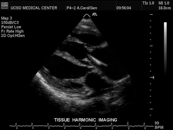
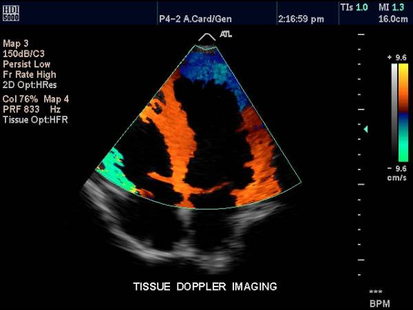
| Septum Using Tissue Doppler Imaging |
| This tissue doppler echo of the heart using a left apical 4-chamber view, and demonstrating a normal heart.
Courtesy Philips Medical Systems 33179 code cardiac heart echo tissue doppler LV RV RA LA |
The diastolic inter ventricular septal (IVS)thickness as measured by echo ranges from 0.7-1.1 cm. Increased IVS thickness is seen early in hypertension and may signify a diastolic abnormality. HOCM which is a genetically determined disease characteristically produces an asymmetrical septal hypertrophy .
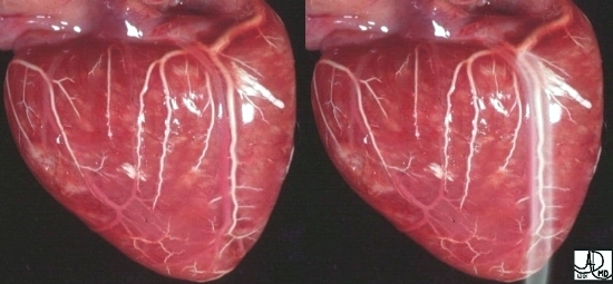
| Change in Septal Size During Systole (right) and Diastole (left) |
| The heart is not static in size and has volume changes every heart beat. The ejection volume is about 70ccs per heart beat. Thus in a normal heart, the diastolic volume of each ventricle is larger than the systolic volume by 70 ccs. In this image of the normal heart, the difference in size is perceptible, with the left image showing contracting ventricles and the right image showing the dilated diastolic ventricles. Courtesy of Philips Medical Systems. 32073 heart normal systole diastole RV LV MV TV LV RV cardiac imaging radiology MRI |
Septal motion during the cardiac cycle is posterior during systole and anterior during diastole. Right ventricular volume overload produces an abnormal systolic interventricular septal motion on echocardiography.
Aging
There is a clinically relevant disproportionate increase in the ventricular septal thickness with age, regardless of gender and in the absence of a history of hypertension. The ratio of ventricular septal to left ventricular free-wall thickness may exceed 1.3 in patients older than 60 years of age, normal being < …”
Parts
LV aspect:
Membranous septum: The fibrous membrane occupying the basal portion of the septum and anteriorly leveled below the mid portion of the right aortic cusp.
Anterior septum (conoventricularis septum, infundibular, supracristal, outlet): A small portion that lies between the anterior LV wall and membranous septum.
Posterior smooth septum(Inlet) : superior 1/3rd to ½ of the muscular part of the septum. It is devoid of trabeculations which differentiate it from the trabecular septum.
Posterior trabeculated septum: Septal wall of the apical ½ to 2/3rd of the ventricle.
RV aspect :
Interventricular portion of the membranous septum: Part of the membranous septum that is overhung by the septal leaflet of the tricuspid valve.
Atrioventricular membranous septum: The part of the membranous septum that lies superior to the tricuspid valve and forms the floor of the right atrium.
Right ventricular Infundibulum : part of the right ventricle which lies between the pulmonary valve above and an imaginary line through the papillary muscle of the conus and upper edge of the membranous septum.
Crista supraventricularis : The crista supraventricularis if oversimplified can be considered synonymous with the infundibular (or conus) ventricular septum.
The appreciation of these terms is essential to understand the various classifications of ventricular septal defects which are discussed in the disease section of the cardiac module.
| Calcification of the LAD |
| This CTscan with contrast through the heart shows heavy calcification of the LAD as well as the circumflex coronary artery as well as calcification of the aortic annulus. Courtesy Ashley Davidoff MD. 15401 code cardiac heart artery coronary calcification calcium calcified CAD atherosclerosis aortic sclerosis annulus imaging radiology CTscan AO aorta |
|
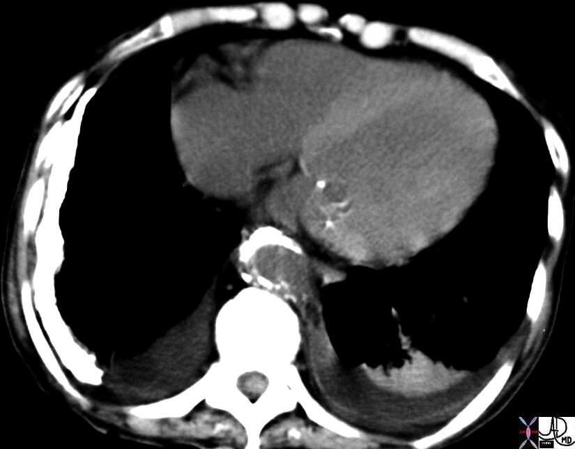
Anemia –Normal Thickness left and Thin (Right |
| 19730 31675b01 heart pleural calcification cardiac interventricular septum IVS LV mitral valve calcification rheumatic heart disease probable chronic hemothorax or empyema anemia fx relatively dense interventricular septum due to anemia CTscan Courtesy Ashley Davidoff MD |
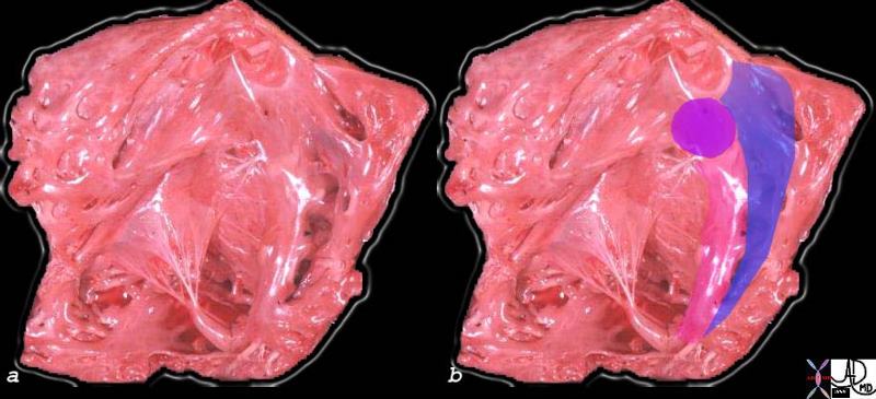
VSD
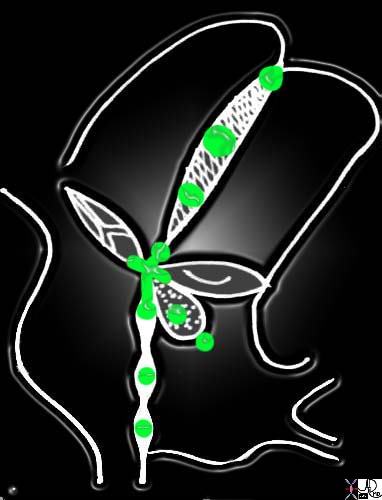
| Defects in the Septa of the Heart |
| 06570e heart cardiac atrial septum ventricular septum IVS sepatal defects ASD of primum type secundum ASD membranous VSD ventricular septal defect muscular VSD VSD of the conal septal conal VSD supracristal VSD subpulmonic VSD ASD of the AV canal type Davidoff art drawing Courtesy Ashley Davidoff MD |
| Membranous VSD |
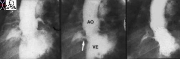
| This is a series of angiograms of the left ventricle in LAO projection showing a puff of contrast into the right ventricle through the interventricular septum. A similar collection of contrast is noted in the the main pulmonary artery just to the left of the aorta. This high VSD is in the position of the membranous septum and thus represents a mebranous ventricular septal defect.. Courtesy Ashley Davidoff MD. 00250 code cardiac heart LV RV VSD membranous MPA aorta AO congenital imaging radiology angiography |
| Muscular Ventricular Septal Defect |
| 10230b01 right ventricle septal band VSD muscular ventricular septal defect congenital heart disease grosspathology Davidoff MD |
| Conoventricular Defect – Single Ventricle |
| 16933 heart cardiac interventricular septum heart dx single ventricle dx ILR dx pulmonary atresia congenital heart disease MRI T1 weighted Courtesy Ashley Davidoff MD |
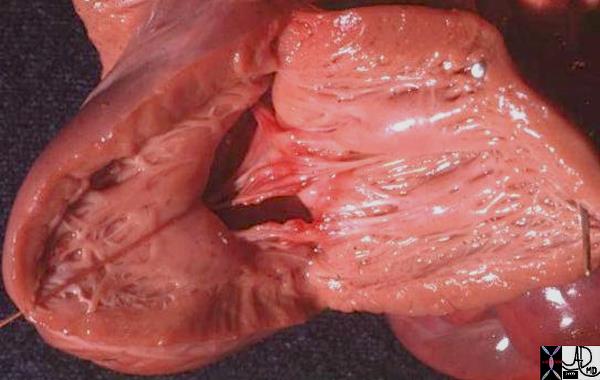
| VSD of the A-V canal type – Complete Endocardial Cushion Defect |
| 01863b01 heart cardiac LV left ventricle IVS interventricular septum mitral valve endocardial cushions complete AVC canal defect cleft mtral valve VSD endocardial cushion defect congenital heart disease grosspathology Courtesy Ashley Davidoff MD |
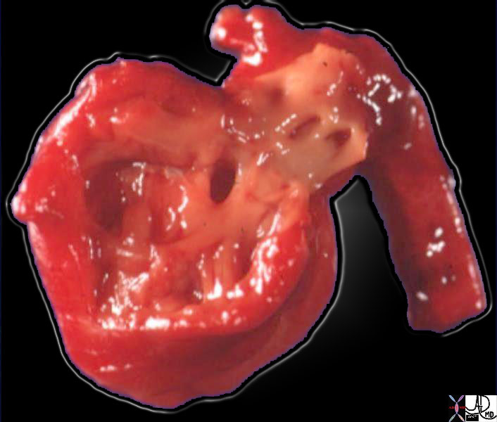
| High VSD |
| 11992b03 heart LV left ventricle interventricular septum ventricular septum VSD ventricular septal defect subaortic grosspathology Courtesy Ashley Davidoff MD |
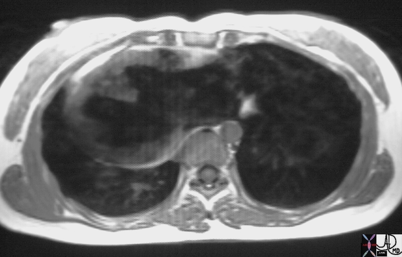
| Abnormal Axis of the Septum – Mesocardia |
| This is a CT scan through the chest in which the apex of the heart points forward. The left ventricle (LV) is left sided and the right ventricle is right sided. There is situs solitus of the atria. This is a case of mesocardia ie neither right nor leftwad pointing apex. Courtesy Ashley Davidoff MD. 19689 code heart mesocardia cardiac imaging radiology CTscan |
| Normal and IHSS |
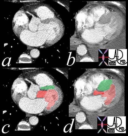
| This series of CTscans shows a normal Left ventricular outflow tract overlayed in bright red in a, with normal septum in green (c) contrasted to a patient with IHSS, where the LVOT is narrowed (b and bright red in d) with a thickened septum with overlay in green (d). Courtesy Ashley DAvidoff MD 39208c01 code cardiac heart CTscan interventricular septum thickened IHSS imaging radiology CTscan |
| Dacron Graft Repair of a Membranous VSD |
| 07932c01 heart LV cardiac defect left ventricle membranous septum ventricular septum interventricular septum VSD dacron graft repair of a membranous VSD papillary muscles grosspathology Courtesy Ashley Davidoff MD |
| Septal and Apical Thinning post MI |
| 26334b heart cardiac interventricular septum IVS apical thinning LV aneurysm apical and septal thinning myocardial infarction CTscan Couresy Ashley Davidoff |
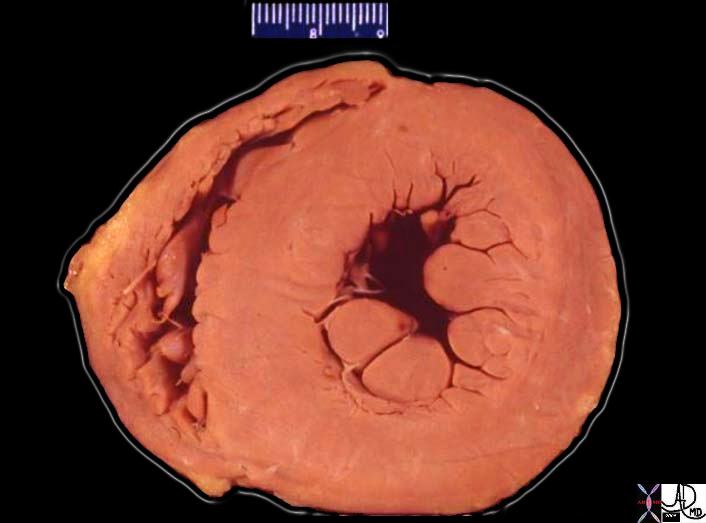
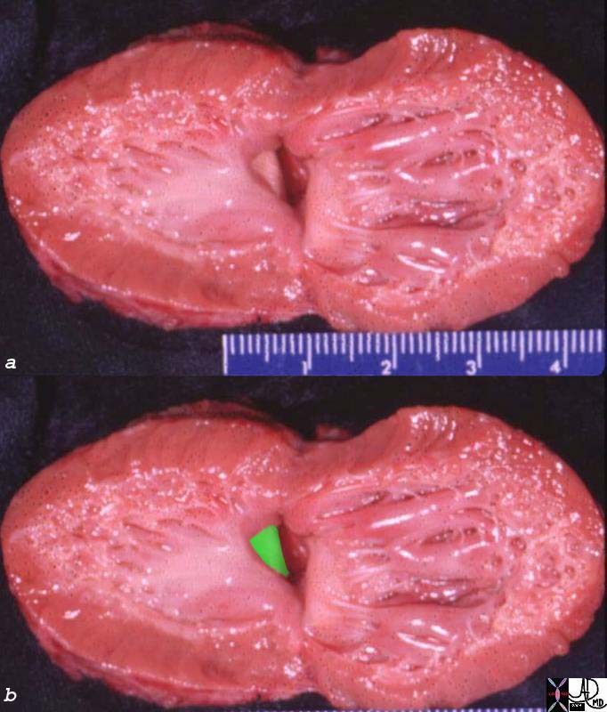
| Concentric Hypertrophy |
| 08028b06 heart cardiac LV left ventricle papillary muscles RV right ventricle LVH left ventricular hypertrophy thickened LV concentric hypertrophy interventricular septum thickened enlarged grosspathology Courtesy Ashley Davidoff MD |
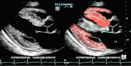
| IHSS Asymmetric Septal Hypertrophy |
| This is a case of focal thickening (bright red overlay) in the region of the septum, which causes obstruction of the LV outflow tract during systole. This condition is known as asymmetric septal hypertrophy – also known as idiopathic hypertrophic sub-aortic stenosis -IHSS. Courtesy of Philips Medical Systems, Ultrasound, and modified by Ashley Davidoff M.D. 32133 |
References
Kitzman D, Edwards WD. Minireview: age-related changes in the anatomy of the normal human heart. J Gerontol Med Sci. 1990;45:M33