
THE CV STRUCTURES VISIBLE ON THE LATERAL EXAM –
Ashley Davidoff MD
RULE OF 1/3 rds
- Normal Anteriorly
- RV takes up 1/3 of retrosternal space ((Sternomanubrial jn to xiphi)
- Inferiorly
- LV takes up 1/3 of the hemidiaphragm
- Posteriorly
- LA 1/3, LV 2/3

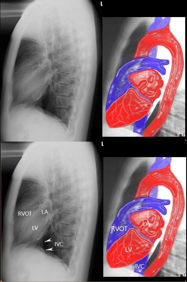
the anterior border of the chest is divided into thirds; 1/3 for the RVOT and 2/3 for the retrosternal air space
the posterior border of the heart is divided into thirds; 1/3 for the LA and 2/3 for the LV.
the diaphragmatic border is divided into thirds; 1/3 for the LV and 2/3 for the rest of the diaphragm
Ashley Davidoff MD
15416C02Wlateral.8 rule of thirds
- Abnormal
-
- Anteriorly
- RVE
- RV > 1/3 of retrosternal space
- RVE
- Posteriorly
- LAE
- LA >1/3 of posterior heart border
- Also elevates left main stem bronchus
- LAE
- Anteriorly
-
 LATERAL EXAMINATION RVE AND LAE – MITRAL STENOSIS PULMONARY HYPERTENSION AND COR BOVINUM
71 year old Asian female with rheumatic heart disease dominated by calcific mitral stenosis mild MR, moderate tricuspid regurgitation
 NORMAL and RVE
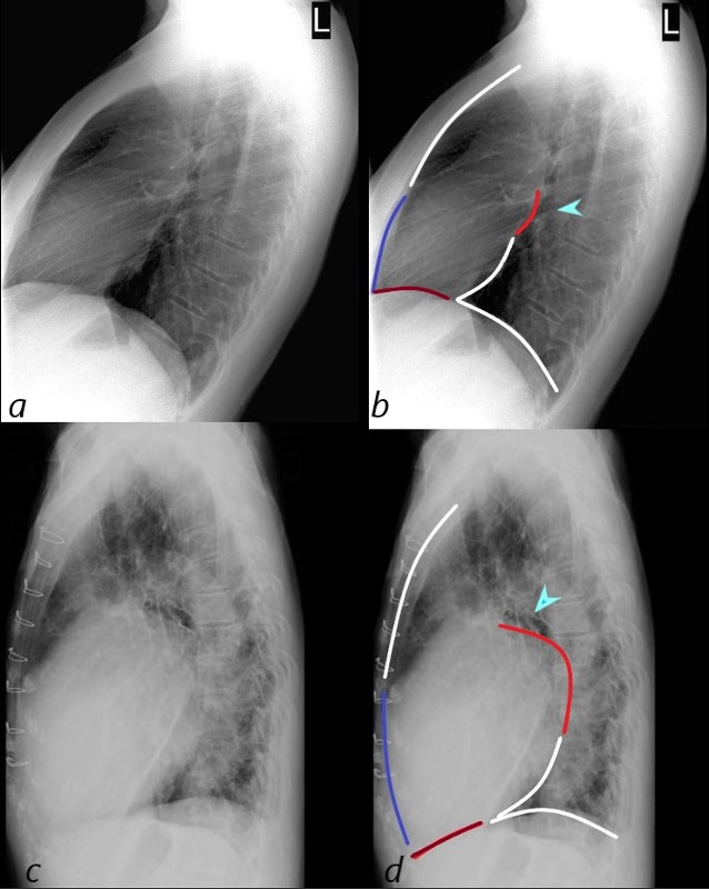
The normal lateral CXR (a,b), shows anterior and superior border of the heart (anterior white arrowhead) occupying 1/3 of the border between the sternomanubrial junction and the diaphragm.
The posterior and inferior white arrowhead shows the posterior border of the heart occupied by the RV taking up 1/3 of the distance of the diaphragm.
Images c and d represent left ventricular enlargement showing that the LV occupies about half the length of the diaphragm, (red arrowhead) while the retrosternal distance is unchanged and normal (white arrowhead).
Ashley Davidoff MD
Left Ventricular Enlargement
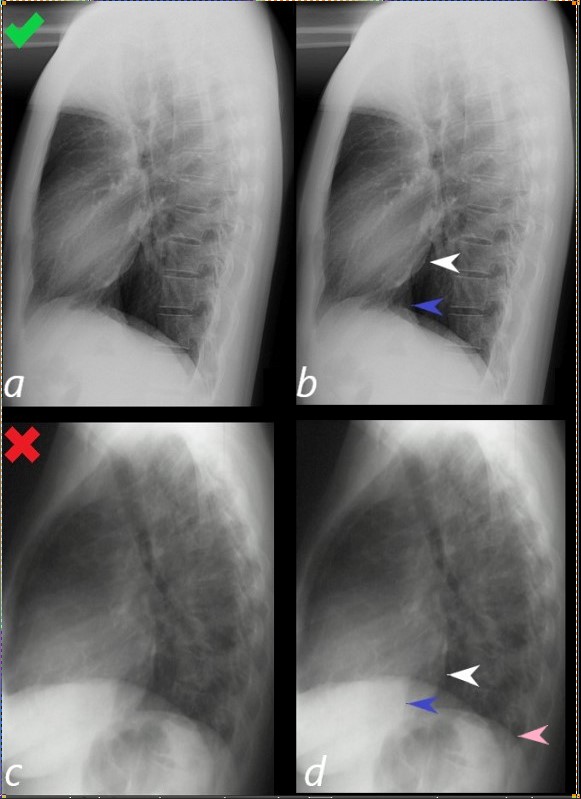
Lateral examination of a chest x-ray (CXR) shows the normal in the upper row (a,b) and the abnormal and enlarged in the bottom row (c,d).
The objective evaluation is based on the relative positioning and size of the LV (white arrowhead) in relation to the IVC, (blue arrowhead), and the left hemidiaphragm (pink arrowhead)
Ashley Davidoff MD
15416C02Wlateral LV01L.8
WHERE IS THE RIGHT ATRIUM?
In the middle of the soft tissues of the heart and so it is not border forming
 NORMAL RIGHT ATRIUM – SAGITTAL MRI WIT SVC AND IVC
- When the RA enlarges-
- it moves laterally and anteriorly see CT below
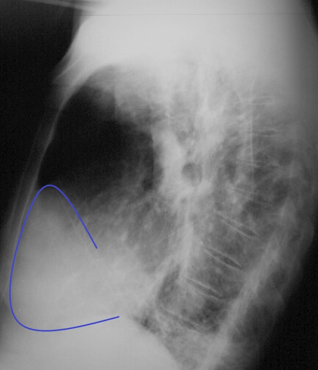
83-year-old male with significantly enlarged right atrium, as well as a mildly enlarged left atrium with evidence of CAD, atrial fibrillation and hypertension.
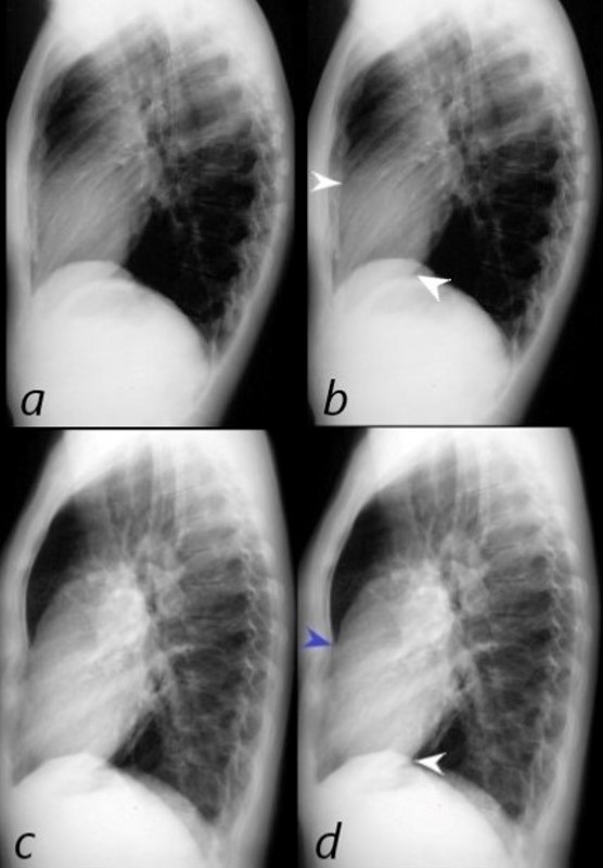
On the lateral CXR, the right atrium the blue line overlay indicates the position of the RV against the anterior chest wall. In addition, the right atrial appendage is so large (see CT below) that it lies against the sternum. The RA therefore occupies the entire retrosternal air space and results in decrease of the retrosternal airspace. This is the rare instance when the RA shares with, but dominates the anterior retrosternal position with the RV.
Ashley Davidoff MD
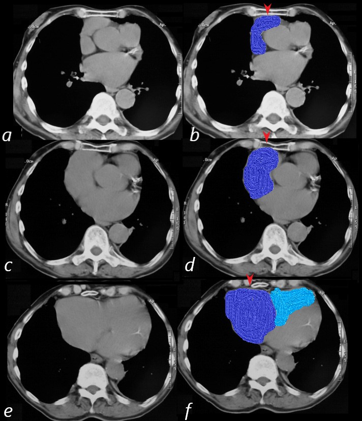
83-year-old male with significantly enlarged right atrium, as well as a mildly enlarged left atrium with evidence of CAD, atrial fibrillation and hypertension.
On CT scan the right atrium and appendage (blue overlay (b,d,f) are positioned against the anterior chest wall and sternum (red arrowheads). On the superior most images (b,d) the RV is not present and only takes up a retro-sternal position alongside the right atrium on the most inferior image (f)
Ashley Davidoff MD