The atrial septum is a fibromuscular wall that separates the right and left atrial chambers.
Structurally it is characterized by two overlapping embryological structures, the left-sided partially fibrous septum primum, and the right-sided muscular septum secundum1.
The mean thickness of the septum primum covering the fossa ovalis was 1.8±0.7 mm. The septum primum was significantly thicker in patients with significant with disease of the atrioventricular valve (2.0±0.6 mm) compared to those without significant disease of the atrioventricular valve (1.6±0.7 mm; p<0.01).
Mean IAS thickness was 2.56 (SD 0.92) mm at anterior region (AR), 1.89 (SD 0.75) mm at fossa ovalis (FO), and 2.95 (SD 0.97) mm at posterior region (PR).(ref)
Functionally in fetal life, it serves to enable flow of blood rich in nutrients from the maternal circulation via placenta and IVC to cross from the right sided circulation to the left so that the brain can be supplied with this blood. Postnatally it serves to uncouple the mixing of deoxygenated and oxygenated blood between the two atrial chambers.
Diseases of the atrial septum are mostly congenital. Patent formaen ovale is seen in 30% of adults and can be the source of systemic emboli from the right sided circulation. Atrial septal defect (ASD) is the most common disorder involving the interatrial septum and is also the most common congenital cardiac abnormality diagnosed in adult life .
Diagnosis of atrial septal disease should be suspected clinically in adults presenting with symptoms of dyspnea, fatigue and palpitations, an flow type ejection murmur, loud P2, and a fixed wide splitting of the second heart sound on auscultation. In ASD secundum the EKG shows a RSR prime pattern suggesting RV overload. The CXR may show an enlarged right ventricle and pulmonary arteries and increased flow. Transthoracic echocardiography allows direct visualization of the defect and can be supplemented by bubble study, Transesophageal echo or cardiac catheterization as needed .
Medical therapy, minimally invasive techniques and surgical options are available depending on the disease process in question.
The nomenclature of the atrial septum is confusing since there are a variety of names that describe the same structures. Sometimes the embryological terms are used and sometimes the anatomical terms are used
Diagnostic elements relate to these diseases and include the clinical evaluation for an atrial septal defect (systolic murmur, split fixed P2, right ventricular enlargment, and pulmonary hypertension later in life) EKG showing enlarged RV, and echocardiography that can document defects in the fossa ovalis.
Treatment is usually surgical.
The atrial septum has an upper middle and lower portions, divided as such for both anatomical, embryological reasons.
The upper portion arises from sinus venosus tissue while the middle portion arises from he muscle itself (mesoderm), while the lower portion arises from the endocardial tissue.
Atrial Septum 01669b04 heart cardiac embryology conal septum line drawing right atrium left atrium left ventricle right ventricle interatrial septum interventricular septum atrioventricular septum crux aortic annulus aorta of the heart embryology anatomy Davidoff drawing Davidoff MD 01667b04 01667b04 01667b06 01667b07 01667b09 01667b14 01669b03
Flap Valve – Septum Primum This diagram shows a coronal section of the atria with right atrium noted in blue on the patients right (our left) and the left atrium in red. The septum primum or flap valve (white) is noted with inferior part attached to the inferior limbic band (green) and the superior aspect lying on the left atrial side behind the superior limbic band (also green) Although not shown the most superior aspect of the septum primum is the foramen ovale. When right atrial pressure is higher than LA pressure, blood will flow from right to left (fetal situation). When LA pressure rises post natally as pulmonary arterial resistance decreases, then higher pressure in the LA causes the valve to close against the superior limbic band and eventually seal except for 30% of the population where a fixed seal does not occur. The unsealed state is called a patent foramen ovale. 07538c06 Davidoff art copyright 2008 all rights reserved
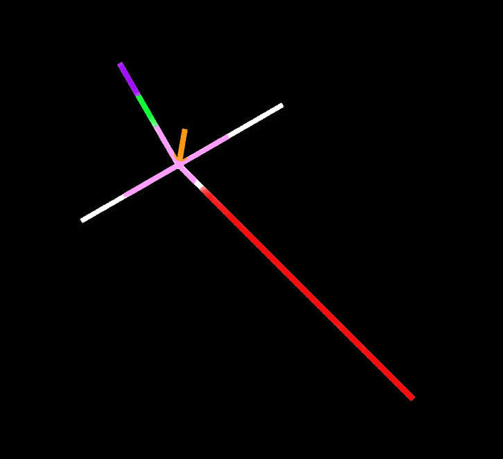
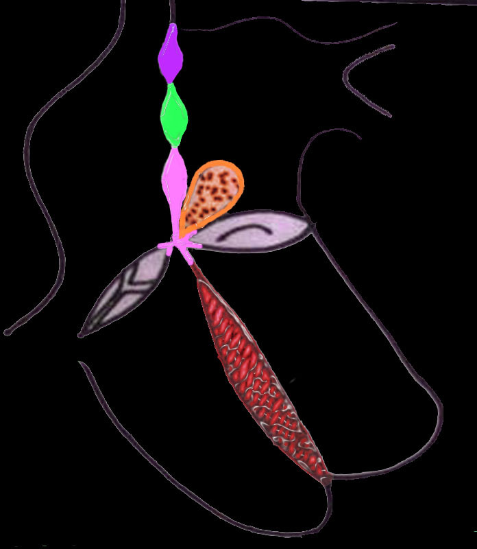
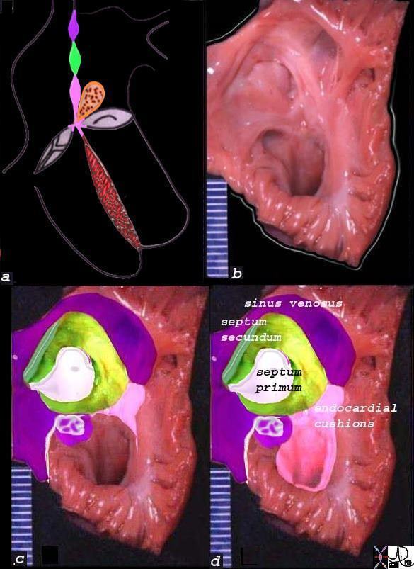
Upper Middle and Lower Septal Regions of the Atrial SeptumThe diagram shows the three portions of the interatrial septum in a with the upper portion (purple deriving from sinus venosus tissue, the middle (grreen) the mesodermal tissue, and the lower (pink) the endocardial cushion tissue. Image b, is an anatomical specimen that is overlaid in reference colors in c, and labelled in d. The middle of the atrial septum is a fibrous membrane called the septum primum, and it is surrounded a rim of muscular tissue called the septum secundum.The septum primum is the middle portion of the interatrial septum and consists of the central solid portion called the septum primum (white) and a hidden foramen ovale which lies on the left atrial side of the septum secundum .01653c11b05a04 Davidoff art copyright 2008 all rights reserved
Septum Primum
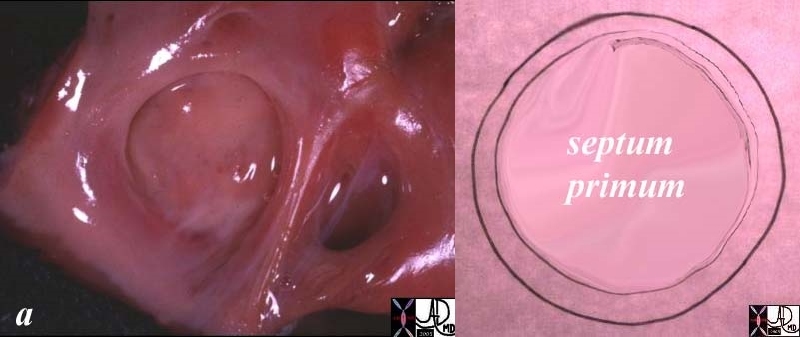
Septum Primum The septum primum is a membrane like structure surrounded by a rim of muscle seen in the first image, and diagrammed in the second.heart cardiac atrial septum septum primum septum secundum sinus venosus right atrium normal anatomy coronary sinus thebesian valve Eustachian valve coronary sinus triangle of Koch atrioventricular node A-V node grosspathology Courtesy Ashley Davidoff MD copyright 2008 all rights reserved 01671c.81s
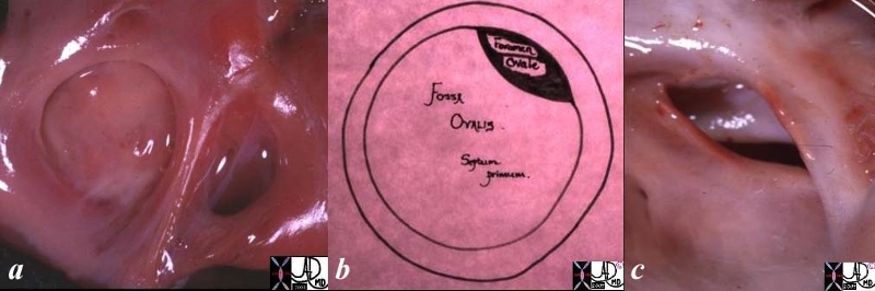
Fossa Ovalis
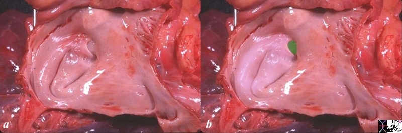
Fossa Ovalis, Septum Primum, and Foramen Ovale The fossa ovalis is the depressed ring like structure surrounded by a rim of muscle seen in the first image, consisting of the septum primum and foramen ovale seen in the second diagram. An anatomical specimen of a patent foramen ovale is seen in the third image characterized by its position and crescentic shape. 01696c01.8s 01671 01670 Davidoff MD copyright 2008 all rights reservedFused Foramen Ovale from the Left Atrial SideThe anatomical specimen shows an opened left atrium with each of the pulmonary veins entering and the left atrial appendage positioned in the upper right hand corner of the image. The septum primum is overlaid in pink, and the characteristic crescent shaped superior border is seen at about 1’oclock on the left atrial side of the septum secundum shown in green. This is an example of a fused foramen ovale.heart left atrium LA pulmonary veins septum primum atrial septum interatrial septum fossa ovalis foramen ovale septum secundum anatomy normal Courtesy Ashley Davidoff MD copyright 2008 all rights reserved 06426c01.8sLeft Atrium – Internal StructureThis anatomical specimen of the opened LA shows the MV, the atrial septum with fossa ovalis, and the laa. The LA is mostly smooth walled except trabeculated finger shaped laa, and bands of the superior limbic band (slb)that surround the fossa ovalis. The oval shaped (black hole) in the fossa is called the foramen ovale which has in this instance been stretched open – better known as a patent foramen ovale.Davidoff MD copyright 2008 Davidoff 32105
Imaging the Septum Primum
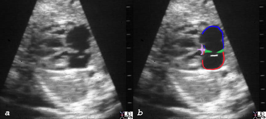
Fetal Ultrasound
The fetal ultrasound shows the right atrium in blue, left atrium in red, limbic band in green and the septum primum white, which is open and lies on the left atrial side.06428c03 heart cardiac atrial septum RA septum secundum superior limbic band inferior limbic band endocardial cushions ventricular septum fossa ovalis fetal echocardiogram LaA left atrium normal anatomy Courtesy Ashley DAvidoff MD copyright 2008
Normal Appearance
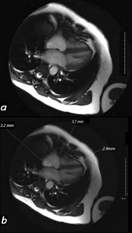
4 chamber of the heart on MRI shows a normal interatrial septum. Three measurements range between 3 to 3.7 mm. The middle portion is the region of the septum primum. Ashley Davidoff MD 130607L
Mean IAS thickness was 2.56 (SD 0.92) mm at anterior region (AR), 1.89 (SD 0.75) mm at fossa ovalis (FO), and 2.95 (SD 0.97) mm at posterior region (PR).(ref)
ASD secundum
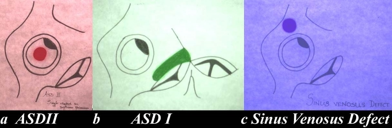
Types of ASD’sThe drawing shows the interatrial septum and the defects associated with each of the components. Image a shows a single defect in the septum primum and this is called an ASD secundum, or a secundum ASD. This is the most common ASD. The second image (b) shows an A-V canal defect in green and it is in the lower portion of the septum and is commonly associated with a cleft mitral valve. The third and least common defect is the sinus venosus defect. (purple)heart inteatrial septum ASD atrial septal defect secundum ASD primum ASD ASD of the sinus venosus type congenital Davidoff art copyright 2008 all rights reserved 01685c02.8
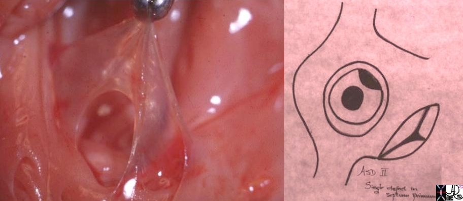
An atrial septal defect of the secundum type is a defect of the septum primum as shown below.
ASD Secundum Defect in the Septum PrimumThe anatomical specimen shows a delicate septum primum held up with a forceps and a hole is noted in the middle characteristic of an ASD secundum. The fosaa ovalis is a wide defect above the septum primum.heart atrial septum ASD secundum septum primum pathology Davidoff art copyright 2008 all rights reserved 01685b01cMRI – ASD SecundumThe MRI shows a flow void due to the turbulence of flow through the ASD secundum – a defect in septum primum.01649b03 Courtesy Ashley Davidoff MD copyright 2008
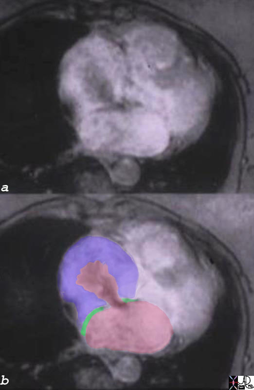
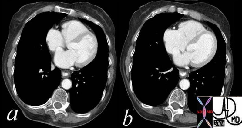
Aneurysm of the Interatrial SeptumThese two axial CT images images through the heart reveal an atrial septum that bulges from the left atrium into the right atrium. The mitral valve is seen in image b, and shows no abnormality. This finding is consistent with a diagnosis of an aneurysm of the atrial septum.Courtesy Ashley Davidoff MD. 38362c01 code CVS heart cardiac LA RA atrial septum aneurysm imaging radiology CTscan 38362c02
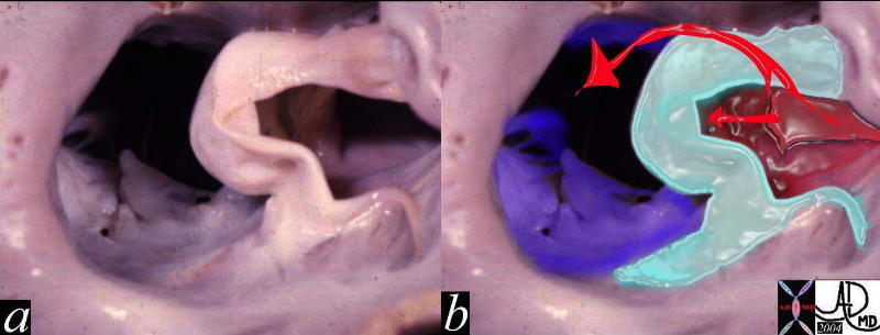
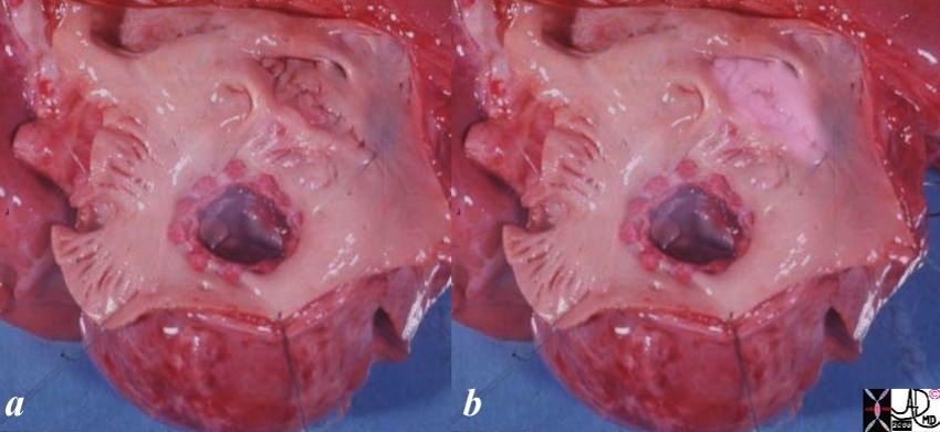
Aneurysm of the Septum Primum in Mitral AtresiaThis is a pathologic specimen of a patient with mitral valve atresia (lime green overlay in b) in this view of the left atrium. The atrial septum shows a redundant perforated septum primum that was restrictive to the high pressure in the LA and an aneurysm resulted. (pale green overlay in b)Courtesy Ashley Davidoff MD. 06829c02 code CVS cardiac heart MV atrial septum ASD aneurysm mitral atresia LA congenital grosspathologyAneurysm of the Septum Primum – Mitral AtresiaThis is a pathological image looking down upon the interatrial septum, showing an aneurysm of the interatrial septum in this patient with mitral atresia and a restrictve ASD. In image b, the light green overlay represents the aneurysm while to the right of the image in red is the left atrium, and to the left of the image (patients right) the tricuspid valve is overlaid in blue. The straight arrow in b reflects the LA blood trying to force its way through the restrictive ASD resulting in progressive bulging of the interatrial septum, while the curved arrow reflects the oxygenated blood that has found it’s way into the RA via a restrictiive ASD.Courtesy Ashley Davidoff MD 06827c01 code cardiac heart RA LA interatrial septum aneurysm ASD septum primum congenital grosspathology
Surgical Repair of an ASD Secundum
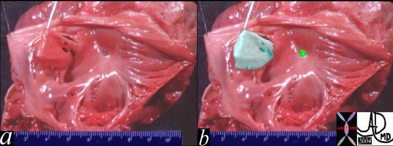
Mitral Valve and ASD Secundum RepairThis anatomical specimen is from an unfortunate pediatric patient who had a mitral valve replaced and an ASD secundum closed. The pink overlay in b shows a suture line opposing the borders of the ASD secundum. A prosthetic mitral valve is noted.heart mitral valve atrial septal defect secundum repair treatment surgery Courtesy Ashley DAvidoff MD copyright 2008 all rights reserved 06704c01.8s
References
Anderson H , Webb S , Brown NA. Clinical anatomy of the Atrial septum with references to its developmental components. Clinical Anatomy 1999;12:362-74.
Anderson H , Brown NA, Webb S. Development and structure of the atrial septum. Heart 2002;88:104-10.
Lazarova D GjorgovNJ. Dimensions of the triangle of Koch. Bratisl Lek Listy 2006;107 (4):107-9.


 Imaging the Septum Primum