
IHSS
Idiopathic Hypertrophic Subaortic Stenosis
Copyright 2008
Ganesh Attaphan MD
Definition
Idiopathic hypertrophic subaortic stenosis (IHSS) is a congenital disorder characterized by marked asymmetrical hypertrophy of the left ventricle, involving in particular the interventricular septum and the left ventricular outflow tract.
Structurally there is hypertrophy of muscles in the left ventricular outflow tract .
Functionally, during systole, the hypertrophied muscle in the outflow tract narrow the subaortic region considerably to produce obstruction to left ventricular ejection.
Clinically patients complain of chest pain , particularly with exercise, dyspnea, syncope and presyncope. Sudden cardiac death can be a presenting symptom of IHSS. On auscultation a crescendo-decrescendo systolic ejection radiating to the suprasternal notch but not to the carotid arteries or neck , best heard between the apex and left sternal border can be heard.
Imaging includes two-dimensional echocardiography is diagnostic for IHSS . Color Doppler flow studies typically reveal associated mitral regurgitation.
Medical and surgical therapies are used to reduce the pressure gradient across the LV outflow tract. Implantation of automatic defibrillators reduces the risk of sudden death .
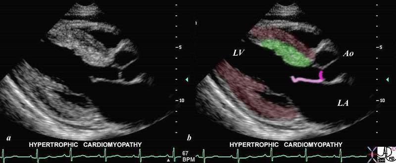
SAM and IHSS |
| This gray scale echo of the heart shows the left ventricle, anterior and posterior leaflets of the mitral valve, the aortic valve and the base of the aorta. There is a focal thickening of the ventricular septum in the left ventricular outflow tract just proximal to the aortic valve. The region is also slightly more echogenic than the remaining myocardium. This case demonstrates a case of asymmetric septal hypertrophy or muscular subaortic stenosis.
Courtesy Philips Medical Systems 33134 code cardiac heart echo LV MV ASH HOCM 33134c04.8s |
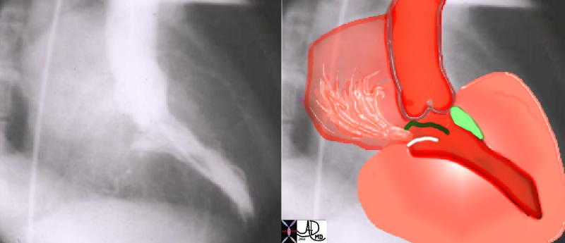
IHSS Angiography |
| This angiogram in RAO projection shows a hypercontractile left ventricle that has a ballet shoe appearance, with mitral regurgitation filling the left atrium. The drawing shows the significant LVH small cavity of the LV, the area of subaortic muscle bundle (green) and the mitral regurgitation caused by the systolic anterior motion of the mitral valve.
Courtesy Ashley Davidoff 34805 cardiac heart MV interventricular septum LVH IHSS SAM MR imaging radiology angiography disease overlay |
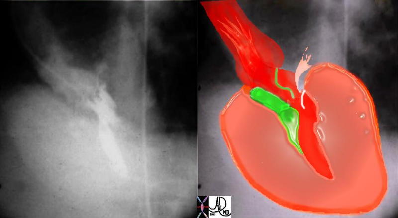
IHSS – RAO Projection |
| This angiogram in LAO projection shows a hypercontractile left ventricle that has a ballet shoe appearance, with mitral regurgitation filling the left atrium. The drawing shows the significant LVH, small cavity of the LV, the area of subaortic muscle bundle (green) and the mitral regurgitation caused by the systolic anterior motion of the mitral valve.
Courtesy Ashley DAvidoff MD 34806 cardiac heart MV interventriclar septum LVH IHSS SAM MR imaging radiology angiography disease overlay |
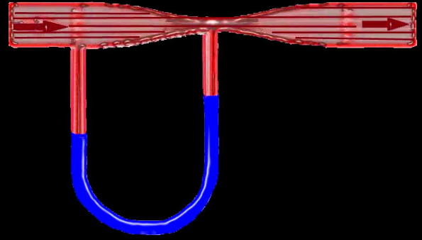
Venturi Effect |
| Flow in this tube is demonstrated by the arrows going from right to left through a narrowing in the tube (red) demonstrating a vacuum or suction effect caused by the sudden acceleration of the fluid as it goes through the narrowing or stenosis. This is seen in the U shaped manometer with the fluid level in blue being relatively higher at the site of the stenosis in relation to the pressure more upstream. The suction phenomenon is known as the Venturi effect is seen in IHSS where the narrowing of the LVOT causes a vacuum effect on the anterior leaflet of the MV resulting in mitral regurgitation. The Venturi effect is also utilised in the functioning of carburettors in fuel driven engines.
34807b05 code heart cardiac SAM IHSS pressure drop vacuum suction acceleration stenosis Venturi effect tube principles |
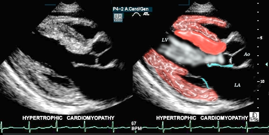
IHSS with Overlay |
| This is a case of focal thickening (bright red overlay) in the region of the septum, which causes obstruction of the LV outflow tract during systole. This condition is known as asymmetric septal hypertrophy – also known as idiopathic hypertrophic sub-aortic stenosis -IHSS.
Courtesy of Philips Medical Systems, Ultrasound, and modified by Ashley Davidoff M.D. 32133 |
References and Links
-
TCV