
Copyright 2009
Introduction
The “twist” in the story is long and complicated, with important implications in congenital heart disease – beyond the scope and need of this anatomy text. In the normal setting we make the following observations: The blue-blooded chambers tend to be rightward, anterior, and slightly superior in position. One of the main structural differences between the right and left sides is that the right ventricle has a separate and distinct outflow chamber – the RVOT or infundibulum. There is an oblique and leftward pointing orientation of the outflow chamber , so that the pulmonary valve lies to the left and superior to its counterpart, the aortic valve. During embryological development, the RVOT, MPA (Main Pulmonary Artery), and aorta twist around each other to result in the structural positioning and relationships of the valves. It always seemed odd that the pulmonary valve coming from the right sided chambers lay to the left of the aortic valve. Under-twisting or over-twisting results in mispositioning of the great vessels in relation to their ventricles with consequent drastic hemodynamic results. Transposition of the great vessels is one such example. The left ventricle arises from the mesodermal germ layer. By the 22nd day of development there is a single heart tube made of an inner endocardial tube and a surrounding myocardial mantle. Starting at the 4th week, the interventricular septum develops to form the distinct left ventricle. This is formed by the inferior endocardial atrioventricular cushion and left conus swelling.
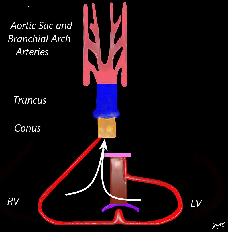
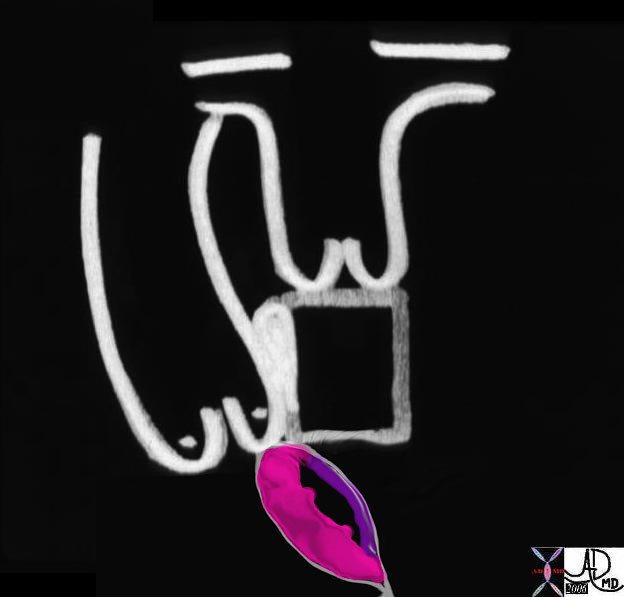
Straight Tube
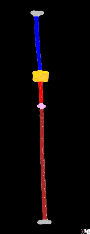
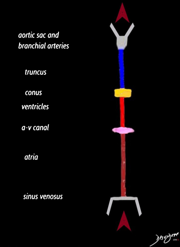
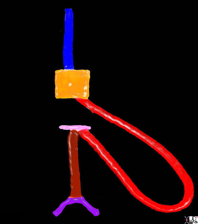
The heart starts as a straight tube. In this stick diagram inflow is through the sinus venosus (maroon) which will evolve into the atria. Blood then passes through the single common atrioventricular valve (short pink horizontal region) which will evolve into the tricuspid and mitral valves. The next vertical limb (red) represents the portion of the tube that will develop into the ventricles. The horizontal orange bar represents the conus – or muscular bundle that lies under the truncus arteriosus. (royal blue). The truncus arteriosus will evolve into the aorta and pulmonary artery.
01477b03 heart cardiac straight tube primitive heart embryology Ashley Davidoff MD
TheCommonVein.net
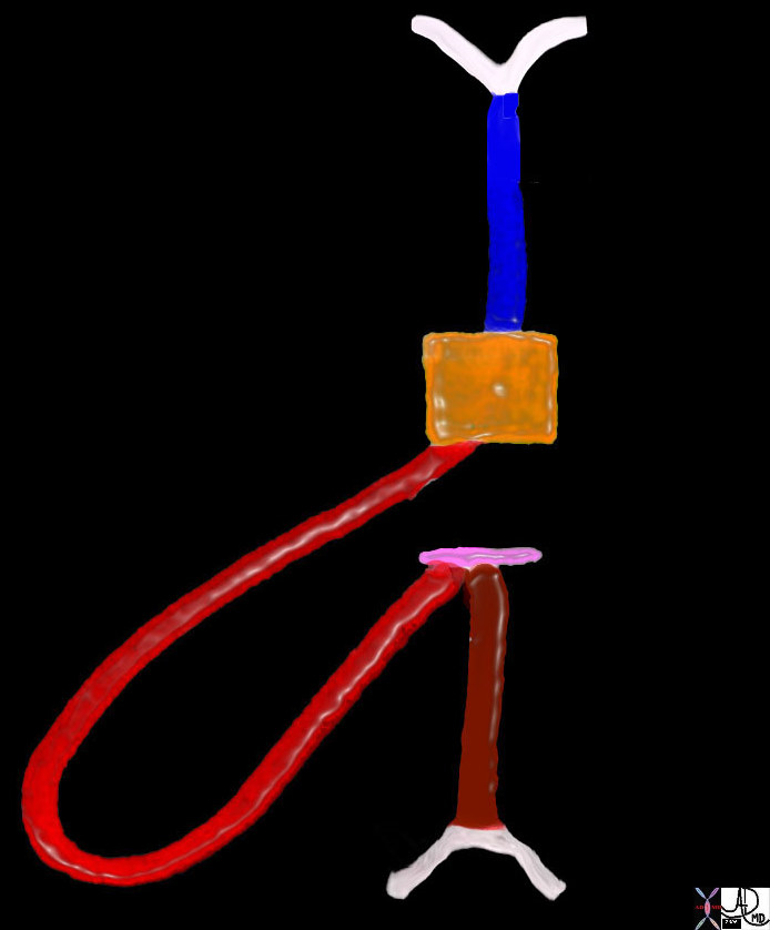
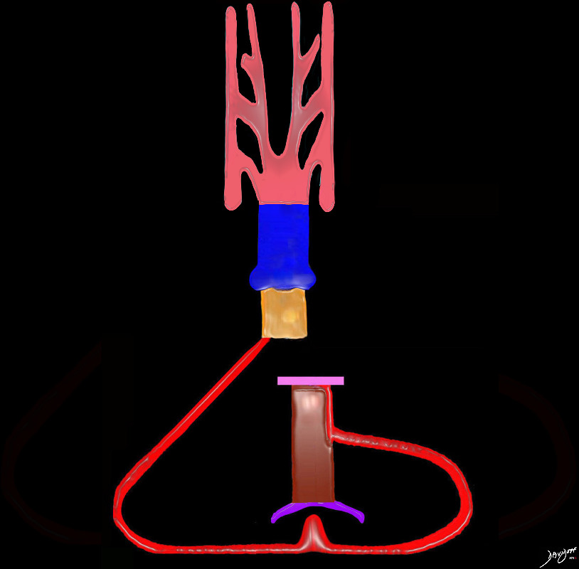
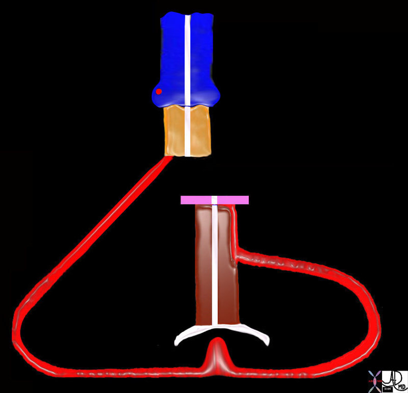
Blood enters the sinus venosus and into the developing atria (maroon), after which it traverses the region of the developing A-V canal (pink) and into the developing ventricles (red) The blood passes through the conus (yellow) and via the truncus (blue)into the aortic sac and branchial arch arteries
Ashley Davidoff MD
TheCommonVein.net
D Loop

Ashley Davidoff MD
TheCommonVein.net
The developing RV in most hearts starts out by looping to the right and also evolves anteriorly because there is room anteriorly in the chest for development since the tube starts out abutting the spine
keywords heart cardiac straight tube d loop D loop primitive heart embryology right ventricle RV sinus venosus atria atrium A-V cushions conus arteriosus truncus arteriosus drawing Ashley Davidoff Davidoff MD 01488b02
DORV phase without Septation
The heart in double outlet right ventricle phase prior to septation, so blood flows into the common ventricular chamber, and out into the conus (infundibulum) and into the truncus prior to entering the aortic sac
Ashley Davidoff MD
TheCommonVein.net
The heart in double outlet right ventricle phase prior to septation, so blood flows intot the common ventricular chamber, and out into the conus (infundibulum) and into the truncus prior to entering the aortic sac
Ashley Davidoff MD
TheCommonVein.net
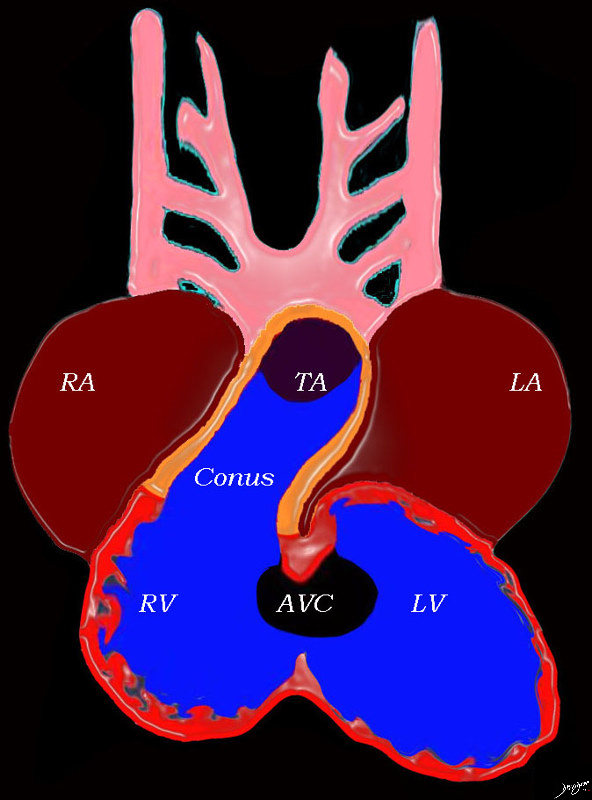
At this stage the heart consists of un-septated atria, ventricles and great vessels, with atria situated posteriorly connected to the ventricles by the AV canal, and the ventricles have a common outlet over the right ventricle to the conus arteriosus, which is connected to the truncus arteriosus
Key words heart cardiac right atrium left atrium right ventricle, left ventricle atrioventricular canal conus arteriosus truncus arteriosus aortic arches RA LA RV LV AVC embryology 01492b04L Ashley Davidoff MD TheCommonVein.net
Septation
Septation occurs at all levels of the heart, including the atria, A-V valves, ventricles, conus, truncal valves, truncus and aortic sac
A D loop will result in positioning the aorta to the right of the pulmonary artery, but at this stage the conus still arises from the developing right ventricle (DORV).
During septation the conus will be divided into a right sided portion that will be subaortic, and a left sided portion that will be subpulmonary
Key words heart cardiac d loop left ventricle is born to the left septation truncus arteriosus conus arteriosus ventricles atrium atria primitive heart embryology 01477e03
Ashley Davidoff MD TheCommonVein.net
TheCommonVein.net
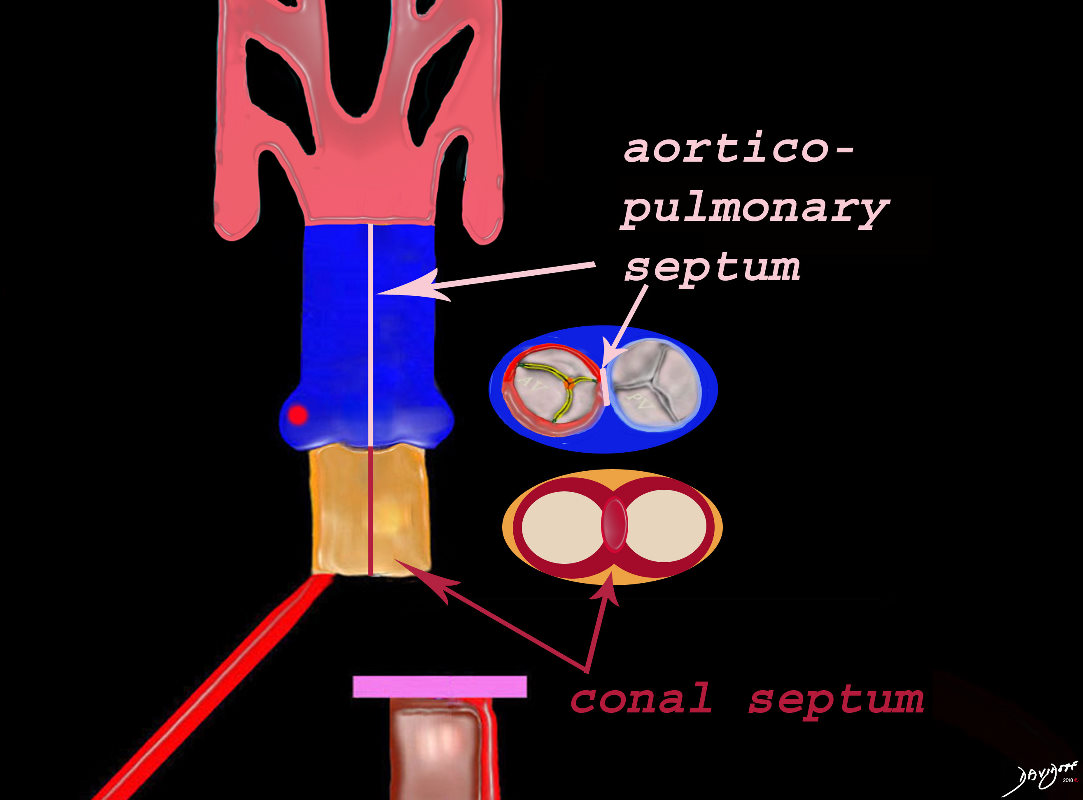
Septa of the Heart
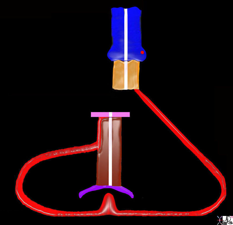
The atrial septum consists of 3 parts; The most superior is the septum derived from the sinus venosus (purple), the middle is the septum primum (green) and the most inferior is derived from the endocardial cushions (pink) The endocardial cushions also contribute to the formation of the of the membranous component of the interventricular septum. The interventricular septum consists of the membranous septum and the muscular septum (red). The great vessels are separated by a muscular conal septum (orange) and the membranous aorticopulmonary septum (light pink).
Ashley Davidoff MD TheCommonVein.net
Septation of the A-V Valves
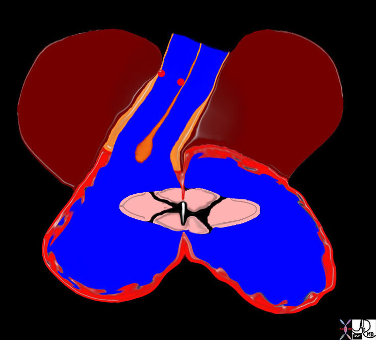
Septation occurs at all levels of the heart, including the atria, A-V valves, ventricles, conus, truncal valves, truncus and aortic sac
The 4 endocardial cushions are separated into 2 evolving valves. On the right side 3 leaflets form – the tricuspid valve and on the left side 2 leaflets of the mitral valve
Key words 01492b12 heart cardiac left atrium right atrium mitral valve tricuspid valve coronary arteries coronary ostia A-V canal atrioventricular canal conus arteriosus truccus arteriosus D loop left ventricle right ventricle embryology RV LV MV TV LA RA aorta pulmonary artery normal drawing Davidoff art Davidoff MD 01492b11 01492b06b 01492b05b01 01492b1201492b14
Ashley Davidoff MD TheCommonVein.net

Septation occurs at all levels of the heart, including the atria, A-V valves, ventricles, conus, truncal valves, truncus and aortic sac
The 4 endocardial cushions are separated into 2 evolving valves. On the right side 3 leaflets form – the tricuspid valve and on the left side 2 leaflets of the mitral valve
Key words heart cardiac left atrium right atrium mitral valve tricuspid valve coronary arteries coronary ostia A-V canal atrioventricular canal conus arteriosus truncus arteriosus D loop left ventricle right ventricle embryology RV LV MV TV LA RA aorta pulmonary artery normal drawing 01492b14
Ashley Davidoff MD TheCommonVein.net
Cono-Truncal Septation
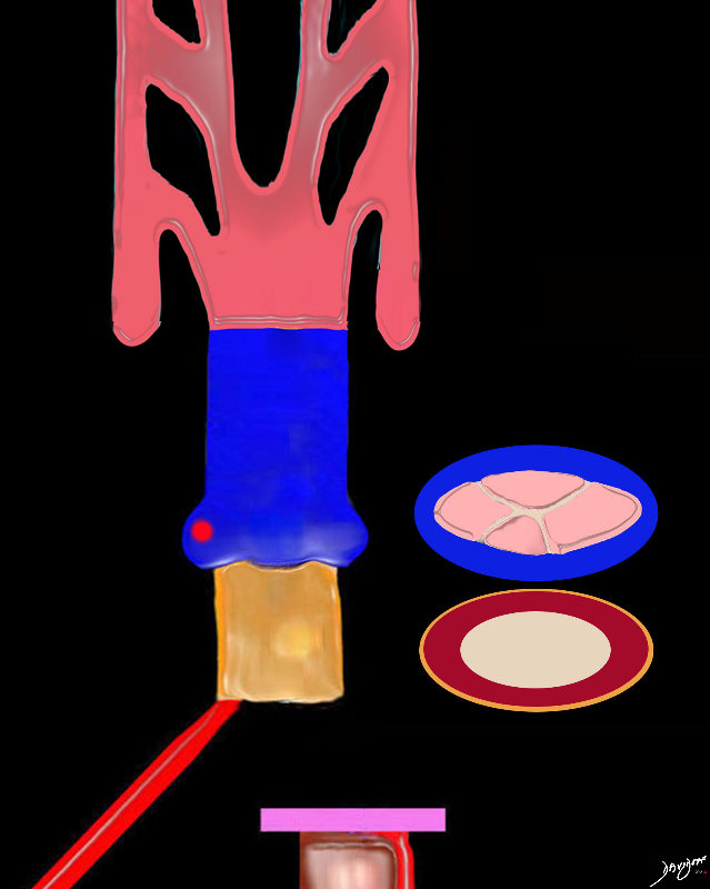
The conus (gold exterior consists of a single muscular tube prior to septation.
Theorigin of the truncus contains 4 truncal cushions which are going to develop into the aortic and pulmonary valve following septation.
Ashley Davidoff MD
TheCommonVein.net
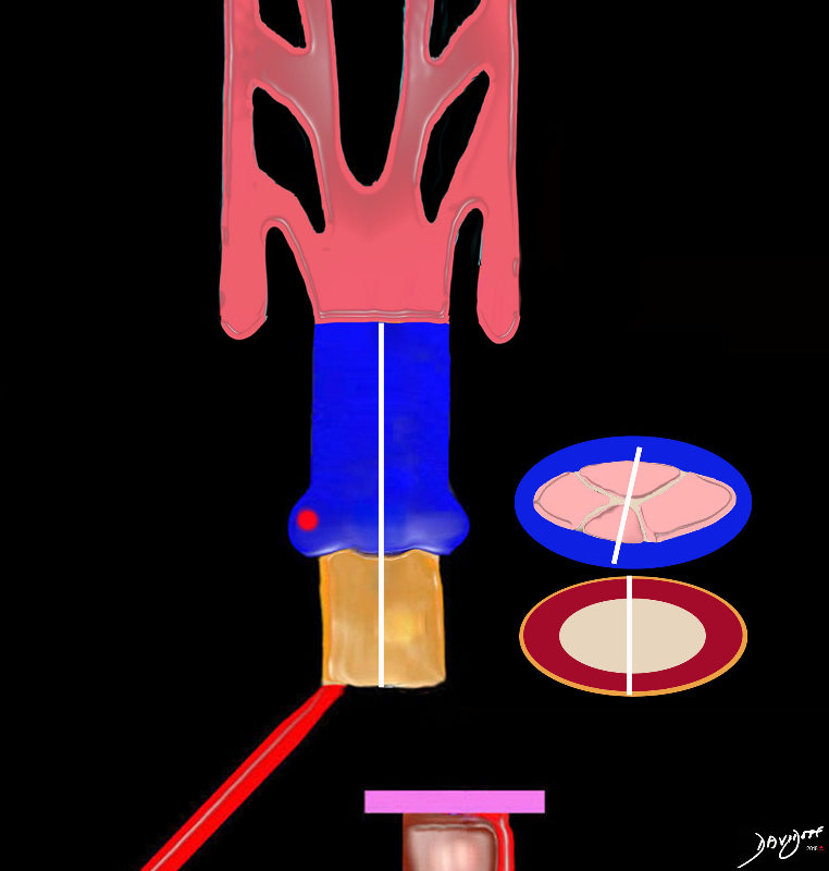
A D loop will result in positioning the aorta to the right of the pulmonary artery, but at this stage the conus still arises from the developing right ventricle (DORV).
The truncus after it divides will have two valves consisting of 3 leaflets each
During septation the conus will be divided into a right side portion that will be subaortic, and a left sided portion that will be subpulmonary
Ashley Davidoff MD
TheCommonVein.net
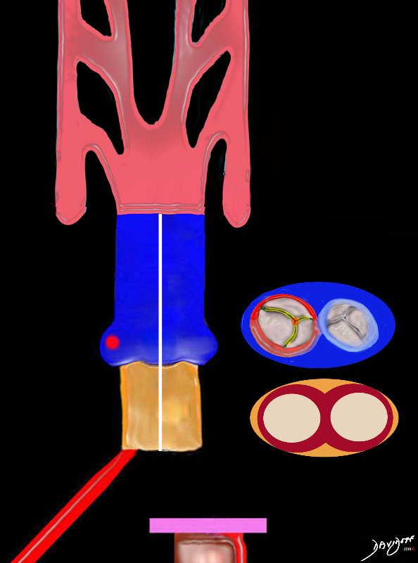
The conus now consists of muscle bould tubes, and the aortic valve and pulmonary valve are well formed. Double outlet right ventricle persistsresulting in both great vessels receiving blood from the single ventricle.
Ashley Davidoff MD
TheCommonVein.net
The conus consists of muscle bound tubes, separated by the conal septum, and the aorta and pulmonary artery are separated by the aorticopulmonary septum. The morphology and physiology are consistent with a double outlet right ventricle state. Both great vessels receive mixed blood from the “single” ventricle.
Ashley Davidoff MD
TheCommonVein.net
Connecting with Growth and Resorption

At this stage the heart is close to totally septating but before this happens the left ventricle and aorta have to connect in order to create a systemic circulation that is separate from the pulmonary circulation. The aorta lies to right of the pulmonary artery and still has a subaortic conus. In order to connect with the left ventricle and mitral valve, the subaortic conus has to resorb. Mr Aorta has a his eyes set on Ms Mitral Valve
Ashley Davidoff MD TheCommonVein.net
2018
01806
Ashley Davidoff MD
TheCommonVein.net

At this stage the heart is close to totally septating but before this happens the left ventricle and aorta have to connect in order to create a systemic circulation that is separate from the pulmonary circulation. The aorta lies to right of the pulmonary artery and still has a subaortic conus. In order to connect with the left ventricle and mitral valve, the subaortic conus has to resorb. Mr Aorta has a his eyes set on Ms Mitral Valve
Ashley Davidoff MD TheCommonVein.net
A Poem of the Love Affair
At this stage the heart is close to totally septating but before this happens the left ventricle and aorta have to connect in order to create a systemic circulation that is separate from the pulmonary circulation. The aorta lies to right of the pulmonary artery and still has a subaortic conus. In order to connect with the left ventricle and mitral valve, the subaortic conus has to resorb. Mr Aorta has a his eyes set on Ms Mitral Valve
Ashley Davidoff MD TheCommonVein.net
Mr Aorta only has eyes for Ms Mitral
How is Mr Aorta going to reach her?
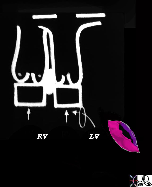
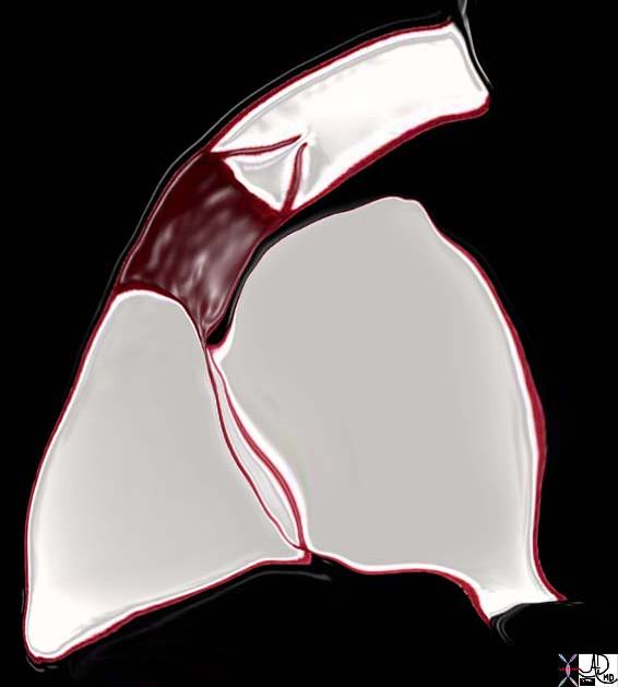
Double Outlet RV Morphology
Ashley Davidoff MD
07427b10
TheCommonVein.net
keywords
06370b04 heart cardiac bilateral conus outflow tract infundibulum aorta pulmonary artery D-loop RV LV cono-ventricular defect bilateral conus atrioventricular endocardial cushion mitral valve anatomy embryology subaortic conus sub-pulmonary conus fibrous continuity mitral valve MV
06370b04
Ashley Davidoff MD
TheCommonVein.net
07427b02
Ashley Davidoff MD
TheCommonVein.net
Ashley Davidoff MD
34814c01.800
TheCommonVein.net
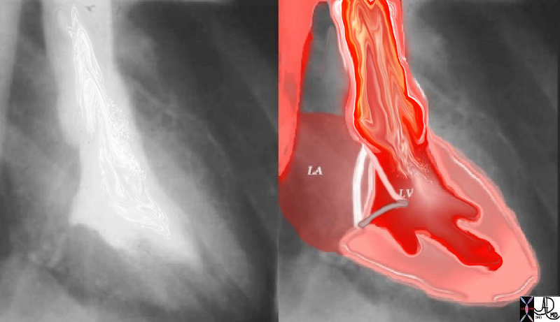
The mitral valve participates in the inflow as well as the outflow of the LV.
The LV does not have an infundibular chamber like the RV.
It is the anterior leaflet of the MV that has this dual function.
The first drawing represents LV diastole, showing the open anterior leaflet acting as the anterior, medial and rightward border of the inflow to the LV.
The second drawing is the systolic phase where this same anterior leaflet acts as the leftward and lateral border of the outflow tract.
Ashley Davidoff MD
TheCommonVein.net
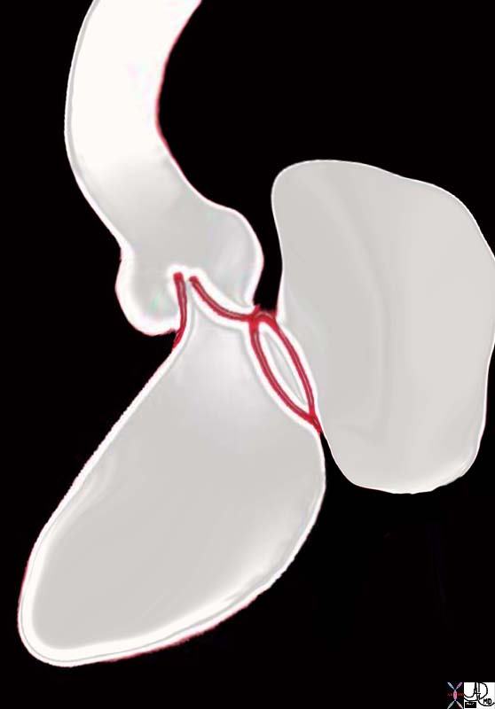
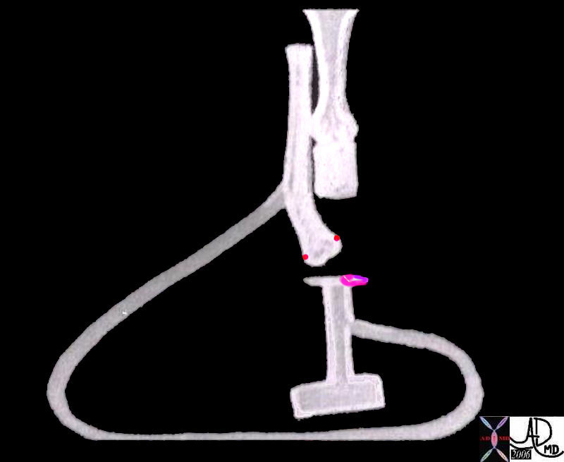
Note how the aorta is tucked facing posteriorly (“heels of the patient”) as a result of the conal resorption
06373b05 heart cardiac aorta mitral valve anatomy embryology resorption of subaortic conus fibrous continuity mitral valve with aortic valve position shape of aorta MV Ashley Davidoff
TheCommonVein.net
The drawing of the right side of the heart in sagittal view shows the two parts of the right ventricle (RV). The triangular inflow portions tarts at the tricuspid valve at the base of the heart, extends toward the pointed apex and then proceeds cranially toward the smooth and tubular right ventricular outflow tract (RVOT) (overlaid in maroon). The RVOT terminates at the pulmonary valve, Note that the valve and RVOT point toward the patients toes. The aorta on the other hand (not shown) points toward the patient’s heels because the subaortic conus has undergone resorption which brings it into fibrous continuity with the mitral valve.
key words
heart cardiac pulmonary artery growth of the subpulmonary conus pulmonary valve infundibulum RV right ventricle RA right atrium IVC inferior vena cava tricuspid valve anatomy embryology position shape of pulmonary artery
Courtesy Ashley Davidoff Davidoff drawing 06376b04 TheCommonVein.net
Keywords infundibulum right ventricle RVOT right ventricular outflow tract aortic valve aortic sclerosis calcification enlarged left atrium heart normal conotruncal relationship cardiac Courtesy Ashley Davidoff MD
31149c.8
Ashley Davidoff MD
TheCommonVein.net
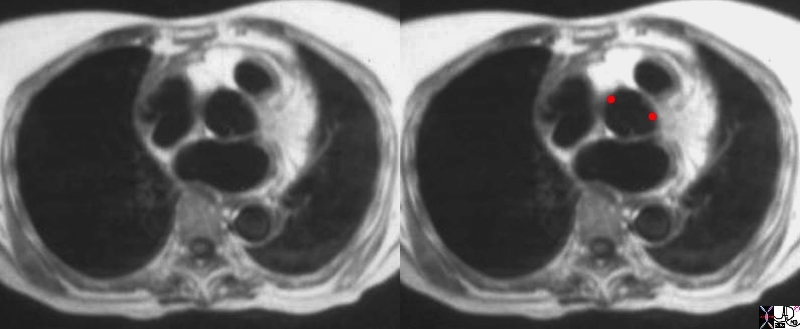
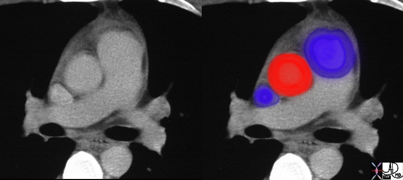
Axial imaging using black blood sequence through the great vessels shows an Ao that is posterior and and to the right and the subpulmonary conus with a rim of muscle positioned anteriorly and to the left.
07523e.8s normal relationship between the aorta and pulmonary artery aorta pulmonary artery conus right ventricular outflow tract MRI T1 weighted
Ashley Davidoff MD
TheCommonVein.net
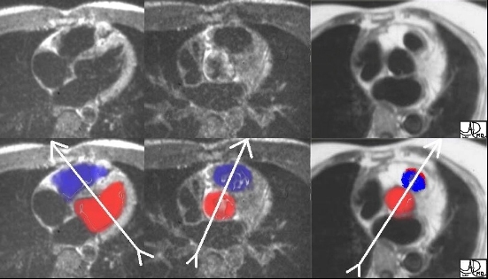
The “twist” story is graphically demonstrated in the following axial MRI images, cutting from inferior to superior as we follow the RV sinus, and RVOT around the aorta. Image 1 shows the triangular RV sinus anterior and to the right of the left ventricle. Image 2 is more superior and shows the RVOT making its way leftward and anterior to the aorta. Image 3 shows the final, distal, and leftward position of the RVOT just below the pulmonary valve. The next set of images with color overlay may be helpful. Ashley Davidoff M.D. 32093 TheCommonVein.net

The aortic valve lies posteriorly and to the right of the pulmonary valve with the final vector directed between 1 and 7 o’clock.
07954eW.800 aorta aortic valve infundibulum RVOT right ventricular outflow tract position relation MRIscan
Ashley Davidoff MD
TheCommonVein.net
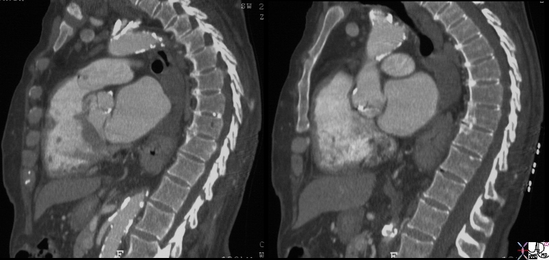
CT showing normal relationship between the aorta (Ao) and pulmonary artery (PA)
CT scan in the axial plane through the great vessels at the level of the pulmonary bifurcation shows that the positional relationship present at the aortic valve level is still maintained at this level. The tubular portion of the ascending Ao is posterior and to the right and the MPA is positioned anteriorly and to the left.
27467c01 aorta pulmonary valve pulmonary artery SVC size position normal CTscan Ashley Davidoff MD
TheCommonVein.net
Applied Embryology in Conotruncal Abnormalities Relating to Growth Regression in the D- Loop Situation
Ashley Davidoff MD
TheCommonVein.net

embryology bilateral conus DORV growth resorption normal mitral to aortic continuity transposition D transposition double outlet right ventricle
Ashley Davidoff art copyright 2019
06394c01L.800s
TheCommonVein.net

Ashley Davidoff MD
TheCommonVein.net
Sometimes the heart, – ie the right ventricle instead of looping to the right will loop to the left. When this happens the aorta will also initially be positioned to the left of the pulmonary artery
Key words heart cardiac straight tube l loop L loop primitive heart embryology right ventricle RV sinus venosus atria atrium A-V cushions conus arteriosus truncus arteriosus drawing
01488b02b01
Ashley Davidoff MD
TheCommonVein.net
Septation occurs at all levels of the heart, including the atria, A-V valves, ventricles, conus, truncal valves, truncus and aortic sac
An L loop will result in positioning the aorta to the left of the pulmonary artery, but at this stage the conus still arises from the developing right ventricle (DORV).
During septation the conus will be divided into a left sided portion that will be subaortic, and a right sided portion that will be subpulmonary
Key words heart cardiac d loop left ventricle is born to the left septation truncus arteriosus conus arteriosus ventricles atrium atria primitive heart embryology 01477e03
Courtesy Ashley Davidoff MD
TheCommonVein.net
embryology bilateral conus DORV growth resorption normal mitral to aortic continuity transposition D transposition double outlet right ventricle
Ashley Davidoff
06394c02L01s
TheCommonVein.net
Summary
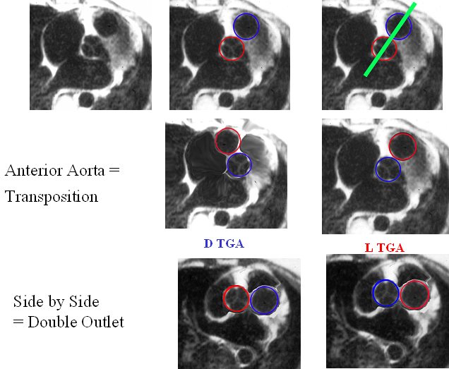
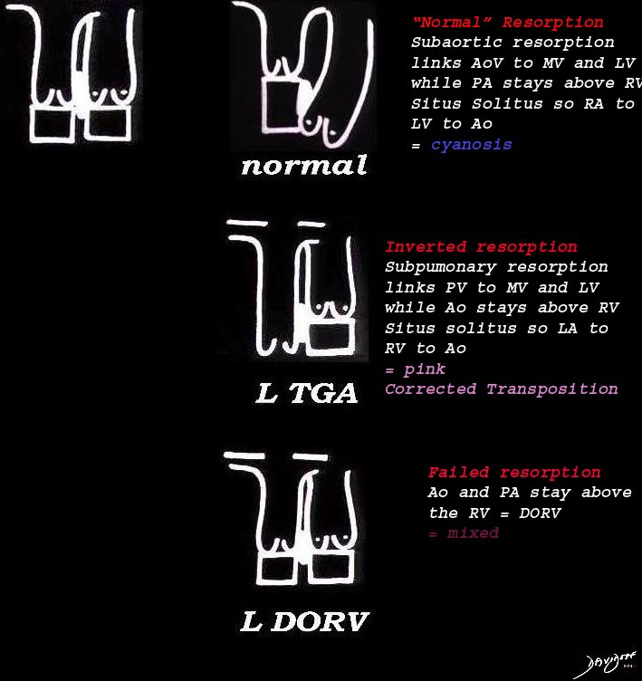
The image reflects the relationship of the aorta and pulmonary arteries in the normal patient, in DTGA, LTGA and DORV. In the normal patient with D loop the aorta (Ao) is posterior and to the right, and the pulmonary artery (PA) is anterior and to the right. In the patient with DTGA, the Ao is anterior and to the right and the PA is posterior and to the left. In an L loop the Ao is anterior and to the left and the PA is posterior and to the right. In double outlet right ventricle (DORV) the great vessels lie side by side and in DORV with a D loop the aorta is to the right and with an L loop the aorta is to the left
86778 01639 01639d02 01639d04 01639f03.jpg Anterior aorta = Transposition
Ashley Davidoff MD
TheCommonVein.net
Powerpoints