
Atrial Septal Defects
Copyright 2008
Introduction
An atrial septal defect is a congenital abnormality of the atrial septum caused by a deficiency in the septum primum, A-V canal, or sinus venosus portions of the atrial septum. A defect in the septum primum results in the most common form called ASD secundum. A defect in the A-V canal portion of the septum results in a ASD primum, while a defect in the sinus venosus is called a sinus venosus ASD. The entity represents about 8-15% of the congenital abnormalities of the heart and it is more common in females. (M:F 1:4) Since pressures in the left atrium are slightly higher than the right atrium there is a left to right shunt in the early phases of the disease. With time, if left untreated, the resulting increased flow to the lungs causes pulmonary hypertension and the shunt reverses, so that a right to left shunt ensues and cyanosis develops. This is called Eisenmenger’s complex.
Diagnosis is suspected clinicaly with a wide fixed splitting of the second heart sound, and a flow murmur across the pulmonary valve. The echo is diagnositic but distinction must be made between a patent foramen ovale (PFO) and the various types of ASDs. The CXR may show rotation of the heart to the left, right heart enlargement, and increased blood flow to the lungs.
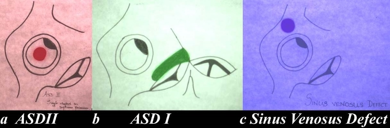
Types of ASD’s |
| The drawing shows the interatrial septum and the defects associated with each of the components. Image a shows a single defect in the septum primum and this is called an ASD secundum, or a secundum ASD. This is the most common ASD. The second image (b) shows an A-V canal defect in green and it is in the lower portion of the septum and is commonly associated with a cleft mitral valve. The third and least common defect is the sinus venosus defect. (purple)
heart inteatrial septum ASD atrial septal defect secundum ASD primum ASD ASD of the sinus venosus type congenital Davidoff art copyright 2008 all rights reserved 01685c02.8 |