
Echocardiography
The echocardiogram provides a real time, safe, accurate, and relatively inexpensive method of evaluating the structures of the heart at any phase of the cycle. Measurements of size including volume, ejection fraction, wall thickness, valve areas, velocity and direction of flow are relatively easy to measure. This technique is the bread and butter of structural cardiac evaluation.
|
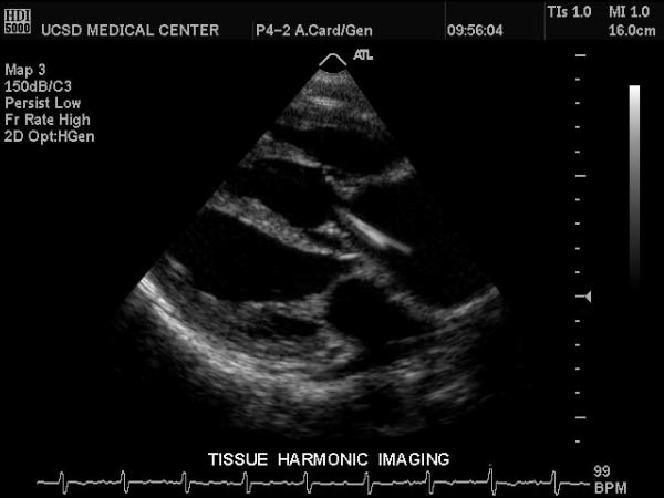
Normal Heart |
| This gray scale echo of the heart showing a long axis of the 4 chambers, and demonstrates normal sized chambers and normal wall thickness.
Courtesy Philips Medical Systems 33186 |
|
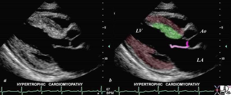
Focal Thickening of the Septum |
| This gray scale echo of the heart shows the left ventricle, anterior (light pink) and posterior leaflets of the mitral valve, the aortic valve (dark pink), and the base of the aorta. There is a focal thickening of the ventricular septum (green) in the left ventricular outflow tract just proximal to the aortic valve. The region is also slightly more echogenic than the remaining myocardium (maroon). This case demonstrates a case of asymmetric septal hypertrophy or muscular subaortic stenosis.
Courtesy Philips Medical Systems 33134 33134c04.8s |
Links and References
Schvartzman et al Normal Values of Echocardiographic Measurements. A Population-Based Study