Patrick J. Lynch, medical illustrator – Patrick J. Lynch, medical illustrator
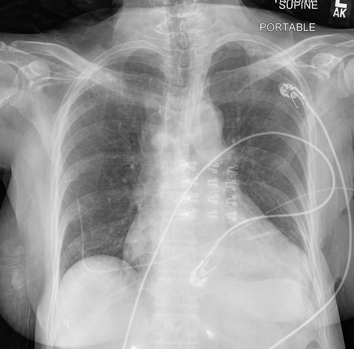
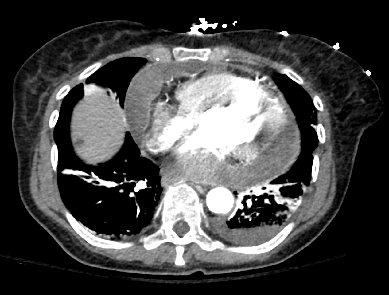
69 year old female
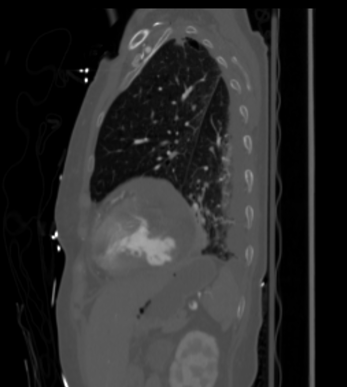
This transverse CT scan through the apex of the left ventricle shows a focal bulge at the apex. There is an associated small focal calcific ation in the myocardium that was noted on a CT 3 years prior in this patient with a diagnosis of a focal pseudoaneurysm of the LV apex. Courtesy Ashley Davidoff MD 33513 heart LV apex calcified pseudoaneurysm inflammation immune rheumatic heart disease RHD cardiac imaging radiology CTscan
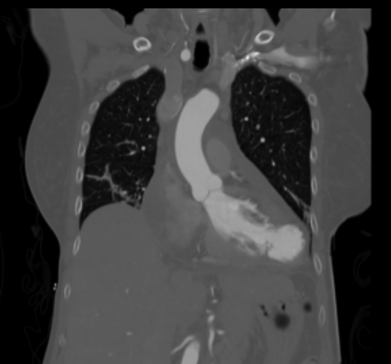
Pseudoaneurysm of the left ventricle, four-chamber echocardiography view