
Definition
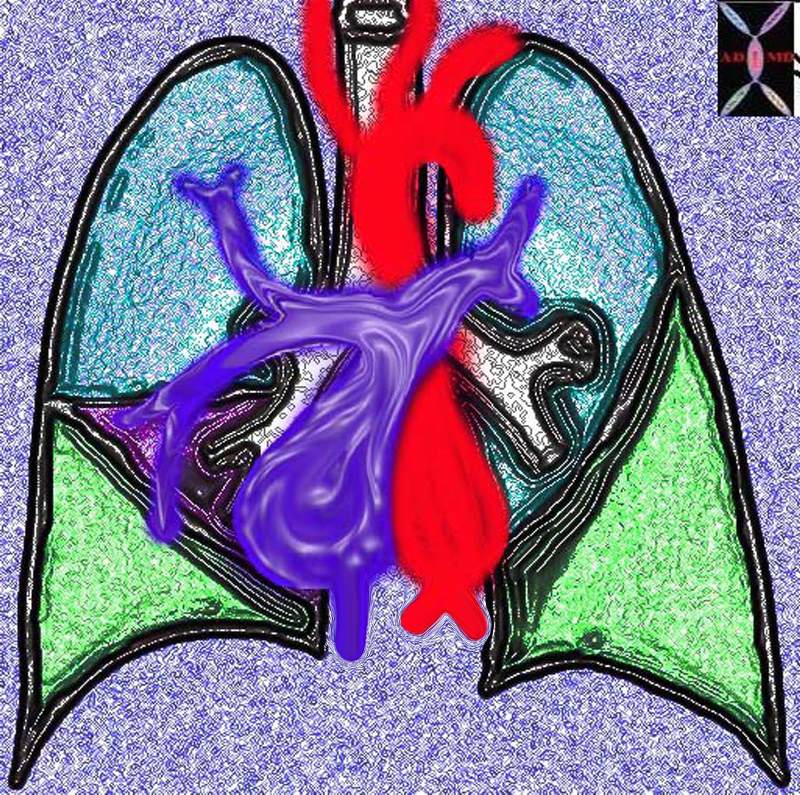
The right ventricle (RV) is the right sided pump of the heart receives deoxygenated blood from the right atrium (RA) via the tricuspid valve and transmits the blood to the lung for oxygenation.
Structurally it is characterized by its position, triangular shape, trabeculated nature, with muscular walls thinner than those on the left . Functionally the RV serves as a pump to transmit blood to the lung.
Sir William Harvey in his De Motu Cordis: described this as “Thus the right ventricle may be said to be made for the sake of transmitting blood through the lungs, not for nourishing them”
Diseases of the right ventricle are usually secondary to LV dysfunction or secondary to lung disease, but may be primary as a result of coronary artery disease, valvular disease, and to less common and rare diseases such as arrhythmogenic right ventricular dysplasia.
The diagnosis of RV dysfunction is suspected clinically by the presence of peripheral dependant edema, ascites, hepatomegaly, an increased jugular venous pressure, which can be increased further by the hepatojugular reflux, a parasternal heave, loud S2(second heart sound) in the pulmonic area and a pan systolic murmur of tricuspid regurgitation .
Imaging includes the use of echocardiography which can reveal enlargement and hypertrophy, usually associated with right atrial enlargement and often accompanied by tricuspid regurgitation.
Medical therapy is used for congestive heart failure or infective endocarditis, and minimally invasive techniques and surgical options are available mostly for intractable tricuspid regurgitation.

davidoff art copyright 2009 Courtesy Ashley Davidoff MD aka 87180b11.8s
aka heart0001
Structural considerations:
Tricuspid Valve and Pulmonary Valve
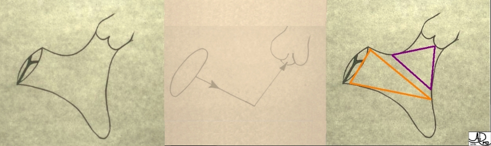
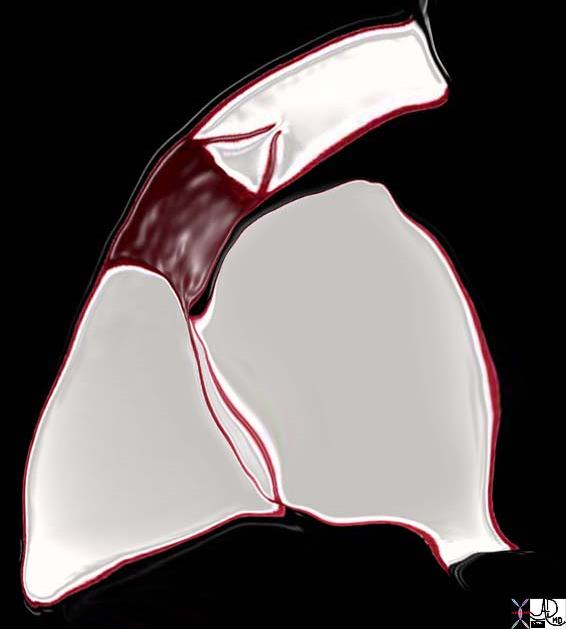
The first diagram is a simple drawing of the right ventricle as seen in a frontal projection. The tricuspid valve is to the right (patient position)and inferior and the pulmonary valve to the left and superior. The arrows of the second diagram show the inflow portion of the right ventricle and the outflow portion. The third and last diagram shows the two chambers that make up the right ventricle. The right ventricular inflow chamber also called the RV sinus is triangular and in orange while the outflow chamber is more tubular or cylindrical and has been called many names – but somehow it does not seem to care. Right ventricular outflow tract (RVOT), and infundibulum seem to be the most popular. Courtesy of Ashley Davidoff M.D. 32087 06610 b trio
Courtesy Ashley Davidoff MD copyright 2019
06925c02.8s
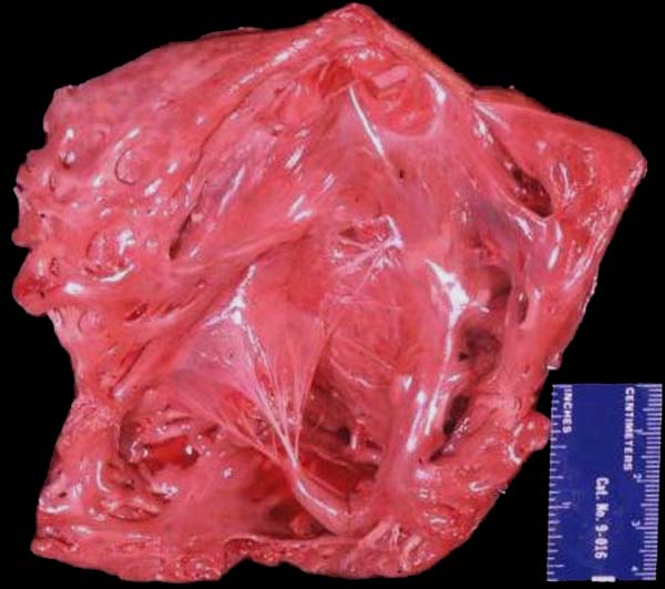
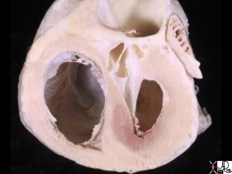
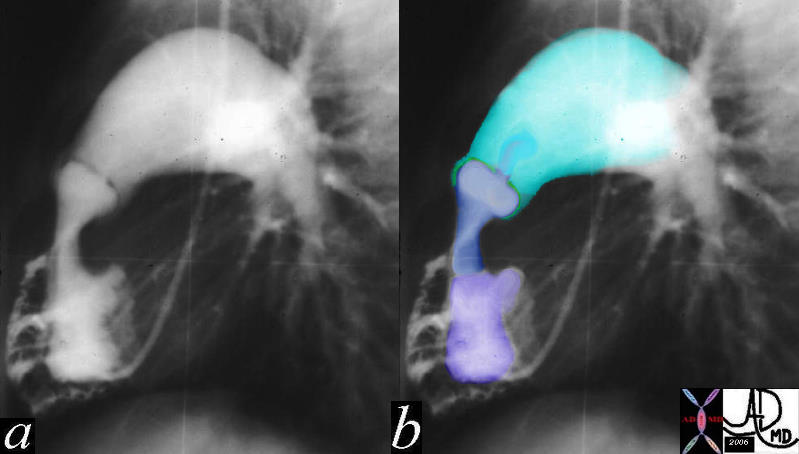
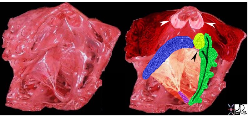
The anatomic specimen shows the appearance of a the inside of a normal right ventricle (RV) with its inflow and outflow (RVOT)
The inflow is identified by the large anterior leafllet of the tricuspid valve (TV) anchored to the septum by the anterolateral papillary muscle. The RVOT is superior to the TV is smooth walled and houses the base of the pulmonary valve note at the superior extent and middle of the RV.
Key words
cardiac heart right ventricle RVOT right ventricular outflow tract parietal band septal band conal septum papillary muscle of Lancisi conal papillary muscle anterolateral papillary muscle pulmonary valve pulmonary artery ventricular septum trabeculae carnii normal anatomy gross anatomy
Ashley Davidoff MD 2019
06409b01
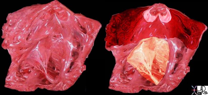
The tricuspid valve is overlaid in yellow showing the large anterior leaflet, and medially placed septal leaflet enabling the identification of the inflow tract and the outflow tract which is overlaid in dark red, The pulmonary valve is overlaid in pink at the superior extent and middle of the RVOT .
cardiac heart right ventricle RVOT right ventricular outflow tract parietal band septal band conal septum papillary muscle of Lancisi conal papillary muscle anterolateral papillary muscle pulmonary valve pulmonary artery ventricular septum trabeculae carneae normal anatomy gross anatomy Ashley Davidoff MD 06409c01
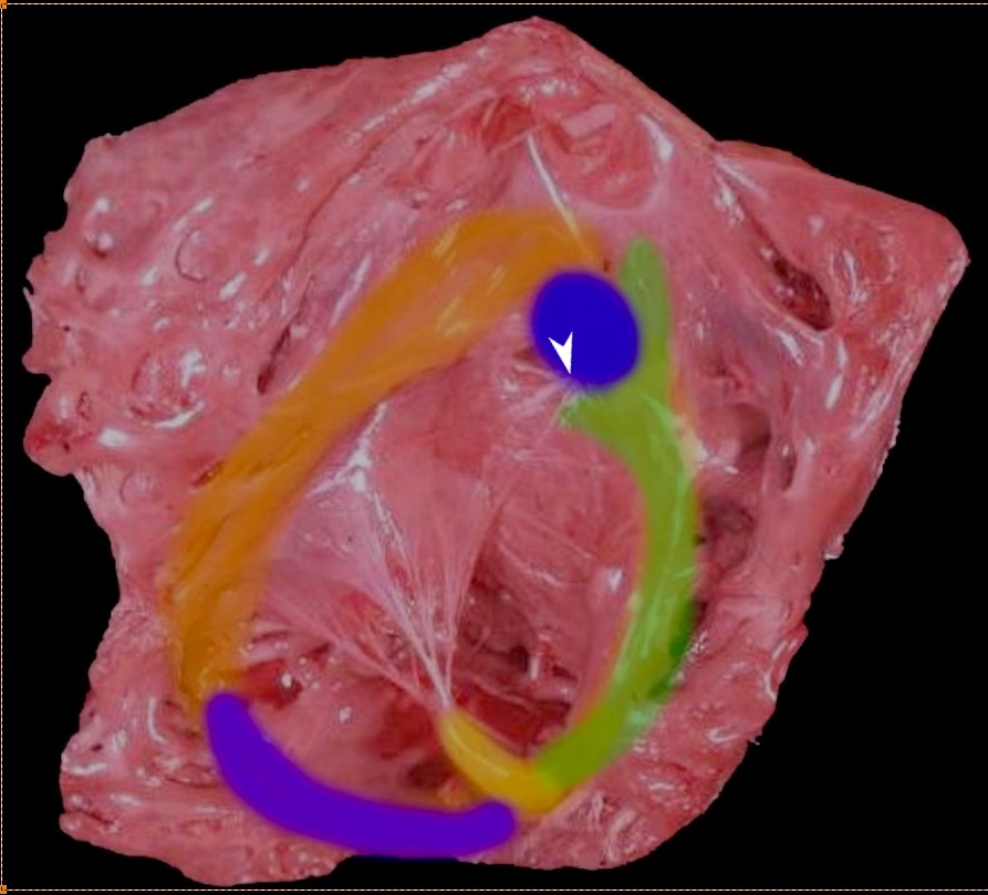
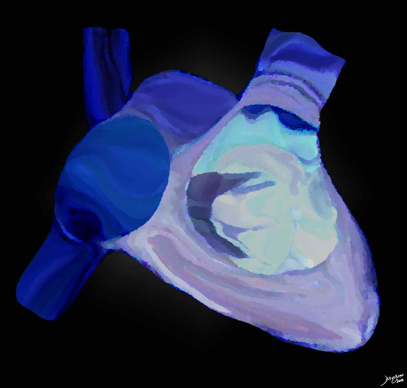
Thr superior aspect of the septal band (green) has two limbs – the “Y” of the septal band. and the conal septum (blue) embeds itself in the Y of the septal band. The (chordae) of the conal papillary muscle (aka papillary muscle of Lancisi- white arrow) also inserts in the “Y ” of the septal band. The anterolateral papillary muscle (yellow) )originates from the septal band) and subtends the anterior leaflet of the tricuspid valve . The moderator band (purple) connects the septum to the the free wall, and the parietal band (orange) completes the muscular ring that separates the inflow from the outflow tract. The pulmonary valve (above the parietal band and conal septum) defines the fborder between the RVOT and conus with the pulmonary artery.
Ashley Davidoff MD
06409 RV muscle bands b01.81
The RV is an asymmetric chamber that appears crescent shaped
|
|
Size
The walls of the RV are muscular with maximum thickness at the base and thinning toward the apex. The free wall thickness of the RV is less than that of the LV with the proportion between them being 1:3, consequent to a low resistance in the pulmonary circulation.
In the mature child and adult, the volume of the RV is larger than the volume of the LV, whereas RV mass is approximately one sixth that of the LV.
|
|
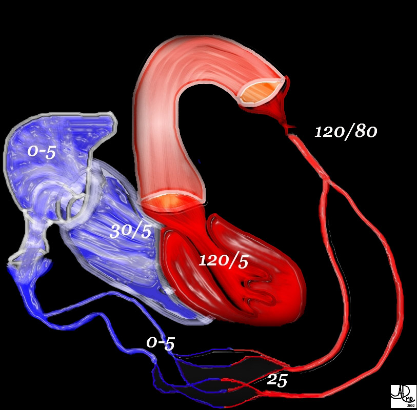
Note the difference in the pressures between the left side of the circulation and the right. The left ventricular pressure reaches a systolic of 120 mmHg, while the right atrial pressure is close to zero. This large difference allows blood to flow through the circulation. pressure principles
49483b01 Davidoff MD Davidoff art
Applied Anatomy
Courtesy of: Ashley Davidoff, MD
aka 37758b01c01.8s
aka heart anatomy P 040
Courtesy of: Ashley Davidoff, M.D heart-anatomy-P-047
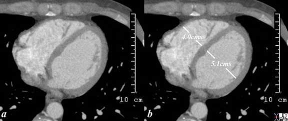
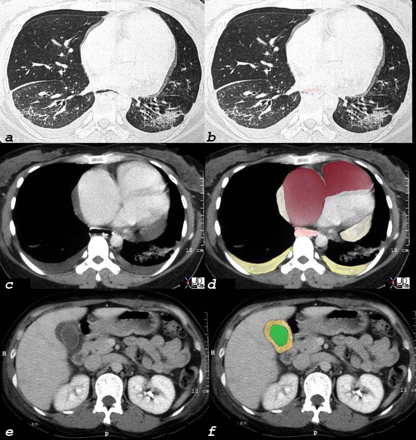
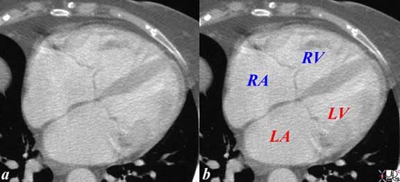
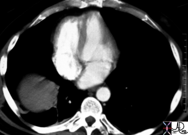
The axial CT scan through the right side of the heart shows an enlarged right atrium and right ventricle in this 88 year old patient with right heart failure with known tricuspid regurgitation The round capaciousness of both chambers provides the subjective impression of RAE and RVE.
key words
cardiac heart CVS RA large enlarged imaging radiology CTscan
Courtesy Ashley Davidoff MD 2019
70344a
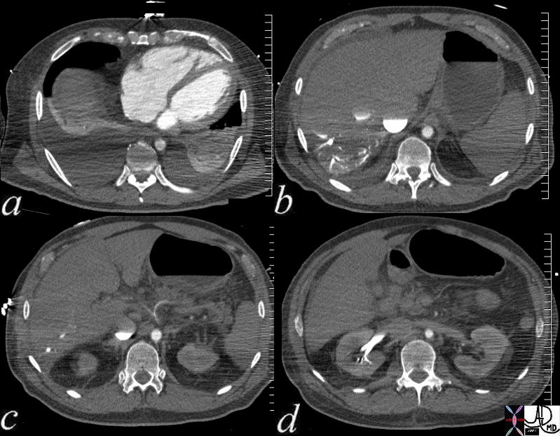
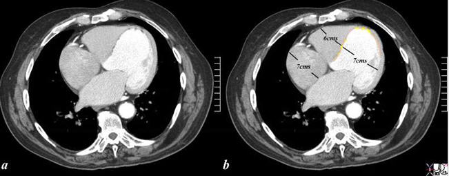
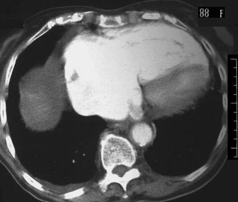
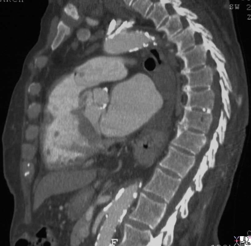
Multiple axial images through the lower chest and upper abdomen in a patient with cardiogenic shock reveals the consequences on the right side of the circulation. In image (a), the right ventricle and right atrium are enlarged and there are bilateral pleural effusions. Image (b) shows stasis of contrast into the IVC with a blood contrast level. The column is relatively static due to peripheral constriction and slow return. There is reflux into the hepatic veins due to tricuspid regurgitation and the reflux extends all the way to the periphery indicating poor forward flow in the hepatic circulation again due to peripheral constriction. Note how small the aorta is due to contraction of the muscular media in this life threatening situation. In image c the celiac axis with branches hepatic artery and splenic artery show severe vasoconstriction. In d the reflux of contrast low pressure extends deep into the renal parenchyma for the reasons outlined above.
key words
heart circulation kidney hepatic vein reflux artery celiac axis spasm aorta small shock CTscan
Courtesy Ashley Davidoff MD 73796c01
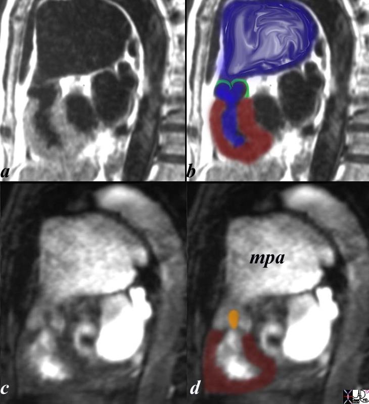
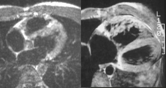
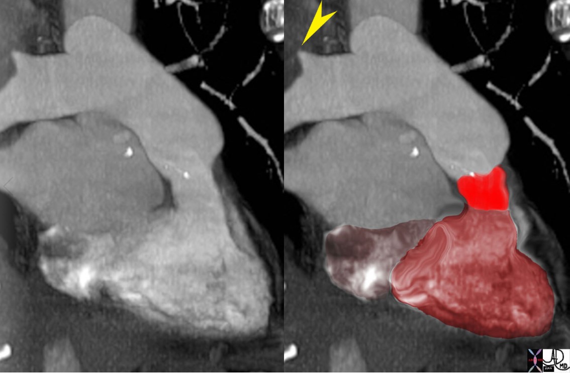
The sagittal view using MRI with black blood (a,b) and white blood(c,d) imaging is from a patient with pulmonary stenosis. The RV (maroon overlay) is hypertrophied, the domed stenotic pulmonary valve (green overlay)is best seen in a and b, and the turbulence through the valve is seen in c, overlaid in orange in d. there is significant post stenotic dilatation of the main pulmonary artery (mpa) inferred by the turbulence in b (blue overlay). code heart cardiac pulmonary valve right ventricle pulmonary artery size enlarged doming stenosis thickened turbulence turbulent flow MRI post stenotic dilatation congenital
Courtesy Ashley Davidoff copyright 2019 15437c03.8s
|
|

Complex Size Changes with Disease
Atrophy Caused by Old Infarction, Swelling with New Infarction, Compensatory Hypertrophy and Normal
This pathological specimen shows a short axis through the heart with the left ventricle (right of image) and the right ventricle (left of the image). The free wall of the left ventricle contains the papillary muscles and is slightly thickened either as compensation to the remaining diseased myocardium or due to other comorbid diseases such as hypertension or aortic stenosis. There is a fresh infarction (black) posteriorly in the left ventricle extending to the posterior wall of the RV as well. The septum is thinned and fibrotic from an old infarction. The anterior wall of the right ventricle is hypertrophied.
Ashley Davidoff MD
15365b01b
aka heart-anatomy-P-031.
Shape
It is is triangular in shape, that in cross section is crescentic

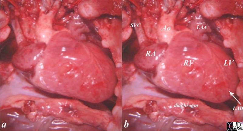
This is an external and frontal view of the heart. The vessel that you see coursing straight down the center of the heart is the left anterior descending artery, (LAD) that runs anterior to the ventricular septum (vertical limb of the cross) separating the right and left ventricles. Conceptualize the triangular shape of the right ventricle, and the ovoid almost football shape of the left ventricle.
Courtesy of Ashley Davidoff M.D. 32078
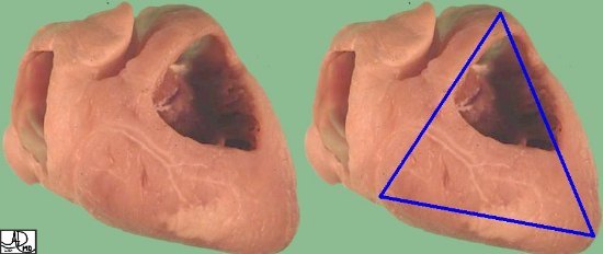
The right ventricle is less sporty in its shape. It is triangular.
(Images courtesy of Ashley Davidoff M.D.32080
key words
heart cardiac pulmonary artery growth of the subpulmonary conus pulmonary valve infundibulum RV right ventricle RA right atrium IVC inferior vena cava tricuspid valve anatomy embryology position shape of pulmonary artery
Courtesy Ashley Davidoff Davidoff drawing 06376b04
key words lung pulmonary artery heart cardiac hypoplastic right ventricular syndrome hypoplastic right ventricle small right ventricle small RV PS pulmonary valve pulmonary stenosis post stenotic dilatation doming pulmonary valve intravasation into coronary arteries suprasystemic RV pressure congenital heart disease size shape RV angiogram angiography Ashley Davidoff MD 15036c01

|
|
|
|
Position
The right ventricle forms the largest part of the anterior surface of the heart , a small part of the diaphragmatic surface and almost entire inferior border of the heart.
|
|
|
|
|
|
|
|
|
|
|
|
Character
Parts
The RV is really composed of two chambers – an inflow or sinus portion and an outflow or infundibular portion. Aristotle had suggested that the heart consisted of 3 chambers. Stella van Praagh a wonderful teacher and friend has translated the original Greek version of Aristotle’s work and firmly believes in her published work that Aristotle had the insight to recognize the right ventricular outflow chamber as the 3rd chamber, with the left and right ventricle each being the other two chambers. It can be divided into basal, mid and apical portions for descriptive reasons. The RV medial wall is shared with the LV and is formed by the interventricular septum, which normally bulges into the RV.
|
|
|
|
It is is triangular in shape, relatively thin walled, but heavily trabeculated, and built to adapt to volume changes rather than pressure changes. The interior of the right ventricle can be described in terms of 3 components: (1)inlet – tricuspid valve , chordae tendineae and papillary muscles, (2) trabeculated apical myocardium (3) smooth outflow tract forming the conus or infundibulum.
|
|
|
|
|
|
Inlet portion of the RV extends from the tricuspid valve to the chordal insertions. The papillary muscles are three in number ( anterior , posterior and septal)
Trabeculated muscular portion is identified by prominent muscle bundles that traverse the chamber from the septum to the free wall. It is this region that, trans-venous pacemaker leads are lodged and biopsy tissues are obtained from.
The right ventricular outflow tract is formed by a smooth collar of myocardium know as the conus or infundibulum.
A C shaped ring of muscle known as the supraventricular crest formed by the parietal band , outlet septum , septal band and moderator band forms the non obstructive opening into the RVOT. The parietal band which separates the tricuspid and pulmonary valves is a free wall structure. The outlet septum separates the two outflow tracts and lies between the left and right commissures of the pulmonary and aortic valve respectively. The septal band is a Y shaped structure with a long broad stem and small inferior and anterior limbs. The two limbs give rise to the septal tricuspid papillary muscle. Apically the septal band merges with the apical trabeculations and gives rise to the to the moderator band that inserts at the base of the anterior papillary muscle.
The membranous septum lies midway between the pulmonary valve annulus and inferior aspect of the tricuspid annulus.
Echocardiography:
A line drawn from the apical to the membranous septum divides the interventricular septum into anterior and inferior portions which in turn can be divided into basal mid and apical portions – giving rise to a total of 6 segments.
The inferior basal and mid segments correspond to the inlet septum.
The apical inferior and anterior along with the mid anterior segments correspond to the muscular septum
The rest of the segments correspond to the outlet septum.
Clinical Considerations:
Differentiation of LV and RV :The RV is defined by its morphology and not by its connections and position. the more apical displacement of the septal leaflet of the tricuspid valve relative to the anterior leaflet of the mitral valve; (2) the presence of a moderator band; (3) the presence of more than 3 papillary muscles; (4)septal papillary attachment of the tricuspid leaflet (5) coarse thicker trabeculations (6) variable locations of the papillary muscles .
The right bundle branch traverses across the septal and moderator bands.
< prev next >