PA CXR
- Enlarged LV
-
- In a Nutshell
- C-T Ratio >.5
- Football in the LV
- In a Nutshell
-
Cardiothoracic Ratio

In this instance images a and c show a ratio that is less than .5 and heart size is considered normal. Images b and d are abnormal since the ratio is greater than .5 and by virtue of the shape of the heart LV enlargement is suggested
Ashley Davidoff MD
Shape Suggesting LV Enlargement
In Clinical Examination LVE?
Down and Out
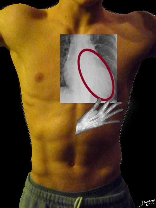 CLINICAL EVALUATION OF LVE – DOWN AND OUT
The left ventricle (LV) enlarges in a downward and lateral direction resulting in the apical impulse displacement and increase forcefulness of the apical tap.
Ashley Davidoff MD
On the AP CXR
LVE =
Down and Out
A Bigger Football
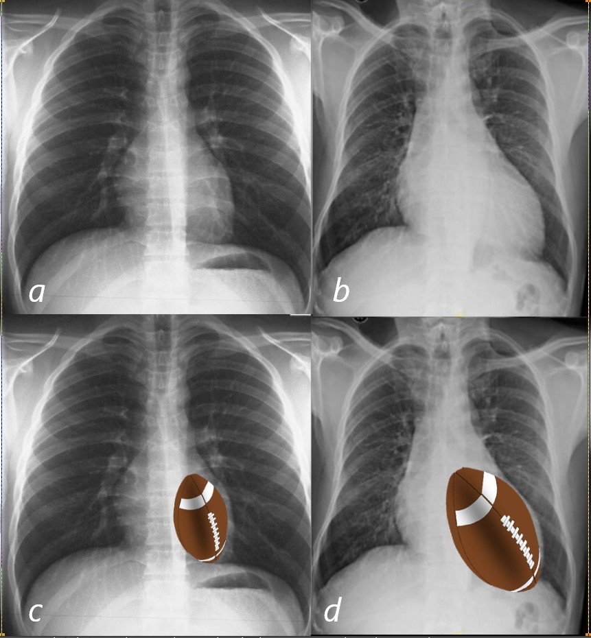
The LV is oval in shape and when it enlarges it advances down and leftward and posteriorly. The oval shape is reminiscent of a rugby ball or a football.
In this instance images a and show a normal shape and size of the left side of the heart . Images b and d are abnormal The heart is enlarged and shows downward and leftward vector
Ashley Davidoff MD
30397 b03L
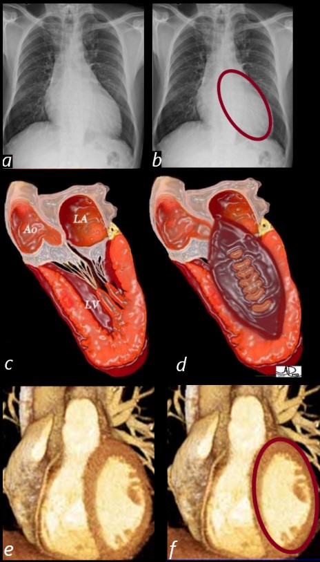
The enlarged LV (a,b) is shaped like an oval and it is likened to a rugby ball or an American football placed on the field at kick off time. LVE on CXR is mostly assessed by an increased cardiothoracic ratio as well as the accentuation of the ovoid shape. (lower images c, d,e, f)
Ashley Davidoff MD
The Lateral – Normal
In a Nutshell
It happens in the backside – LV is the buttocks of the heart
LV is bottom and back
Rule of thirds
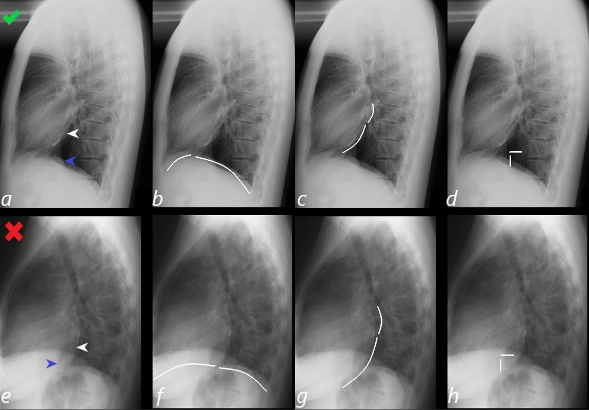
Anatomy of the Normal Lateral Examination
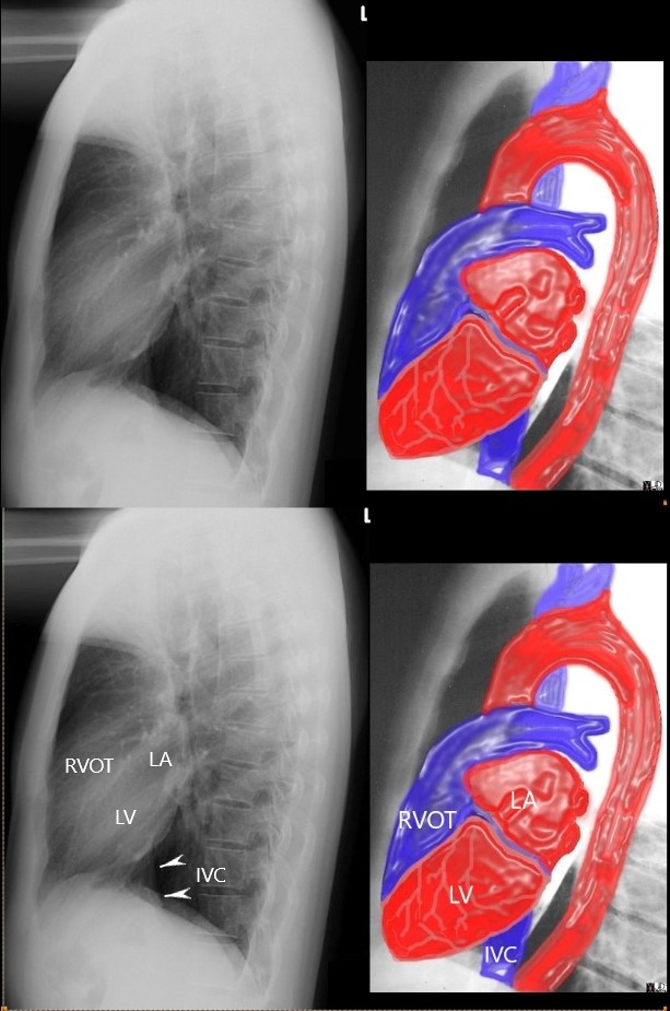
Ashley Davidoff MD
Rule of Thirds
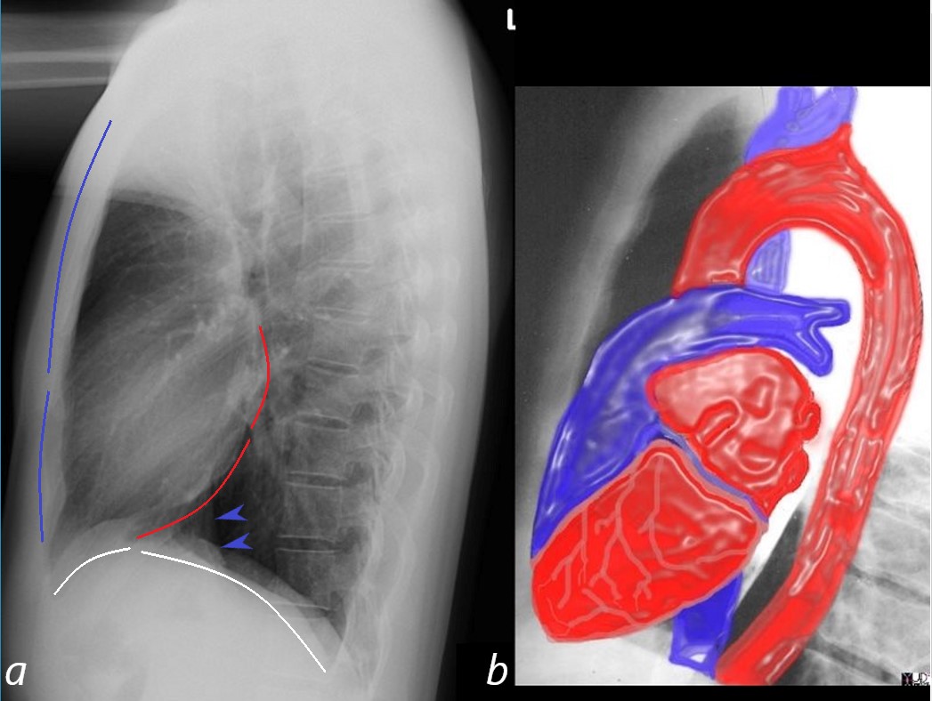
A normal lateral examination of the chest X-ray (CXR, a) shows the relative size and ratios of the anterior border of the heart in relation to the sternomanubrial junction (blue lines), the inferior border of the heart in relation to the diaphragm (white lines) and the posterior border of the heart (red lines)
The first rule of thirds states that the ratio or relative length of the anterior border of the heart (bottom blue line) is about 1/3 t of the length from the inferior border of the heart to the sternomanubrial junction (top blue line) . When the right ventricle is enlarged, the heart takes up more than a 1/3 of this space, and fills in the retrosternal air space indicating right ventricular enlargement RVE).
The second rule of thirds (white lines) states that the ratio or relative length of the inferior border of the heart (front white arc) is about 1/3 the length from the posterior inferior border of the heart to the insertion point of the left hemidiaphragm posteriorly (posterior white arc). When this ratio increases then there is left ventricular enlargement (LVE).
The third rule of thirds (red arcs) states that the ratio or relative length of the posterior border of the left atrium (LA top red arc)) is about 1/3 of the length from the inferior border of the LA to the inferior border of the LV on the left hemidiaphragm (bottom red arc). When this ratio increases then there is left ventricular enlargement (LVE).
A fourth rule (not shown) relates to the relative position of the IVC (blue arrow heads) states that if one draws a 2.5cms vertical line from point where the IVC emerges from the abdomen, and then measures 2.5cms posteriorly, then the posterior border of the LV should be within that 2.5 cms. horizontal line. If the border of the LV is beyond that horizontal line, then LVE is present.Ashley Davidoff MD
15416C02W lateral.thirds
Left Ventricular Enlargement
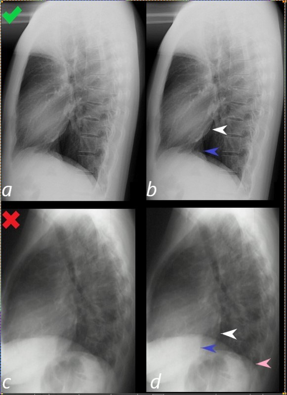
Lateral examination of a chest x-ray (CXR) shows the normal in the upper row (a,b) and the abnormal and enlarged in the bottom row (c,d).
The objective evaluation is based on the relative positioning and size of the LV (white arrowhead) in relation to the IVC, (blue arrowhead), and the left hemidiaphragm (pink arrowhead)
Ashley Davidoff MD
15416C02Wlateral LV01L.8