
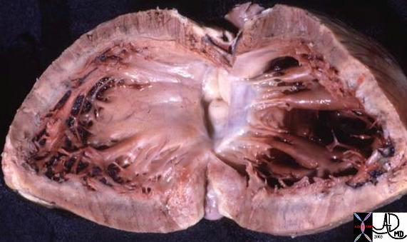
Normal LVOT |
| This anatomic specimen shows the LV with septal side to the patients right and free wall side to the patients left. Note the relatively smooth walled septal surface and the papillary muscle apparatus attached to the free wall side. There are two groups of papillary muscles neither of which are attached to the septum. The anterior leaflet of the MV is in full view and note that it is in fibrous continuity with the left and non coronary cusps of the aortic valve.
Courtesy Ashley Davidoff MD 01817 |

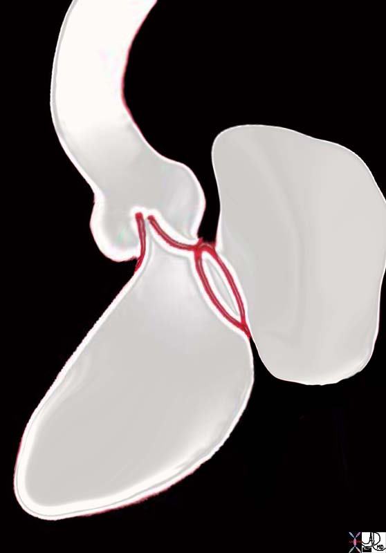
The Mitral and Aortic Valve in Concert |
| ThThe diagram shows how the anterior leaflet of the mitral valve acts as both the infow and outflow of the left ventricle. During systole (left image) the anterior leaflet is forced shut, and forms the left border of the left ventricular outflow tract while the aortic valve is forced open. During diastole (right image) the anterior leaflet of the mitral is open, and acts as the rightward border of the LV inflow tract, while the aortic leaflets are distended with blood closing the aortic valve.
32119d04 aorta aortic valve systole diastole semilunar valves sinus of Valsalva mitral valve left ventricle MV LV normal physiology coaptation of leaflets competence competent Davidoff art Courtesy Ashley Davidoff MD |
Lateral View of the Close Proximity of the Aortic and Mitral Complexes |
| 06373b05 heart cardiac aorta mitral valve anatomy embryology resorption of subaortic conus fibrous continuity mitral valve with aortic valve position shape of aorta MV Courtesy Ashley Davidoff Davidoff drawing |
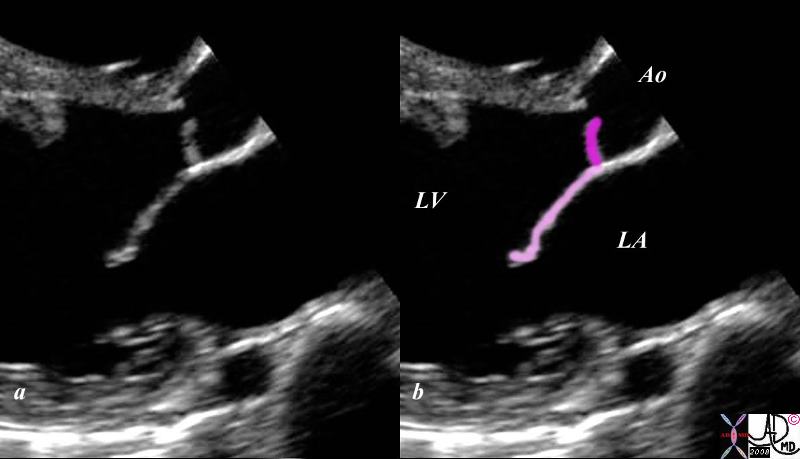
The Normal Mitral Aortic Complex |
| This gray scale echo of the heart shows the left ventricle, anterior and posterior leaflets of the mitral valve, the aortic valve and the base of the aorta. The echo demonstrates the intimate anatomic and physiological relationship of the anterior leaflet of the mitral valve (light pink) and the left aortic cusp(dark pink)..
( Courtesy Philips Medical Systems 33132 code cardiac heart echo LV MV aorta mitral aortic complex normal anatomy 33132c02.8s |

Aortic Regurgitation and the Austin Flint Murmur |
| This gray scale echo of the heart showing a right parasternal long-axis left ventricular outflow view, and demonstrating prolapse of the aortic valve. The patient has a diagnosis of aortic regurgitation. The regurgitant jet of the aortic incompetence created by the prolapsing valve causes the mitral leaflet to flutter..
Courtesy Philips Medical Systems 33124 cardiac heart echo AOV prolapse MV imaging echo 33124c02.8s |
To understand the embryological, anatomical, pathological, clinical and radiological link between the aortic valve and mitral valve
The aortic and mitral valve are linked after the subaortic conus resorbs bringing the valves together in fibrous union. The anterior leaflet of the mitral valve is in intimate contact thereafter with the left and non coronary sinuses. Disease of the one is commonly associated with the other due to their close proximity. Clinically regurgitant changes of the aortic valve lead to changes on the mitral valve and disorders such as ASH, causes mitral valve disease to occur.
Knowledge of the embryology reflects on the anatomy and they both have relevance to the clinical and imaging aspects of these two valves provides an integrated, in depth knowledge for members of a multidisciplinary team in one comprehensive source. Each of the disciplines is treated separately and integrated into the whole module. The viewer will learn the aspects of cellular makeup, histology, anatomy, pathology, diseases, imaging, and clinical disciplines. The addition of historical events, art, and culture of the brain adds richness to the program.