
Structural Parts
- Muscular Elements
- Anterior Wall
- Apex
- Inferior Wall
- Lateral Wall
- Posterior Wall
- Base
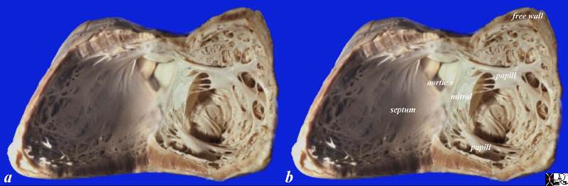
This normal anatomical specimen has been sectioned in long axis along the ventricular septum; then opened like a book with the septal wall seen on the left side of the image, and the free wall with the pair of papillary muscles seen on the right of the image. Note the fibrous continuity of the anterior leaflet of the mitral valve with the cusps of the aortic valve..
code 15389c05.8s Courtesy Ashley DAvidoff MD copyright 2009
Base of the Heart
A Closer Look at the Crux of the Heart, Membranous Portion of the Ventricular Septum, Mitral Valve to Aortic Valve Fibrous Continuity
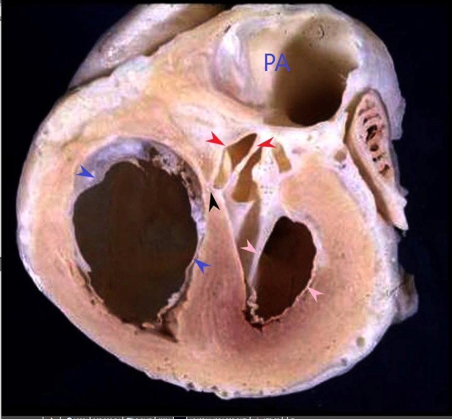
The anatomic specimen shows the right ventricle and the tricuspid valve (blue arrows), the left ventricle and the mitral valve (pink arrows) , with fibrous continuity with the aortic valve, and the region of the membranous septum (black arrow) at the crux of the heart
Ashley Davidoff MD
Mitral Valve
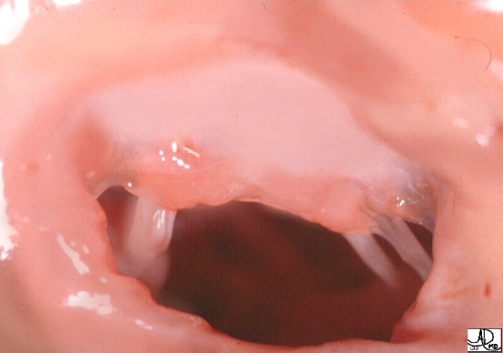 Anterior Leaflet of the Mitral Valve
The anatomic specimen taken from the left atrium through the mitral annulus shows the broad anterior leaflet of the mitral valve ..
Courtesy of Ashley Davidoff M.D. code cardiac heart normal MV mitral valve anatomy 32106

Leaflets of the Mitral Valve
The mitral valve consists of two leaflets; an anterior shield shaped leaflet that extends deep into the LV, and a posterior leaflet that occupies a greater percentage of the circumference of the annulus, but is more shallow in its extension into LV.
code cardiac heart normal MV mitral valve anatomy Courtesy of Ashley Davidoff M.D.32116
Inflow and Outflow Portion of the LV and the MV
There is an inflow and an outflow part of the LV. In diastole the mitral valve (MV) is open and the inflow portion receives atrial blood
07999 Ashley Davidoff

07992 Ashley Davidoff
Applied Anatomy
Mike Tyson and the Right Upper Cut
Right Sided Structures Anterior and Inferior
Left Posterior and Inferior

Right Sided Structures Right Sided, Anterior and Inferior, Except the Pulmonary Artery which is Leftward and Superior
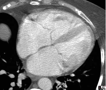
Septum Apex and free Wall in Axial Projection
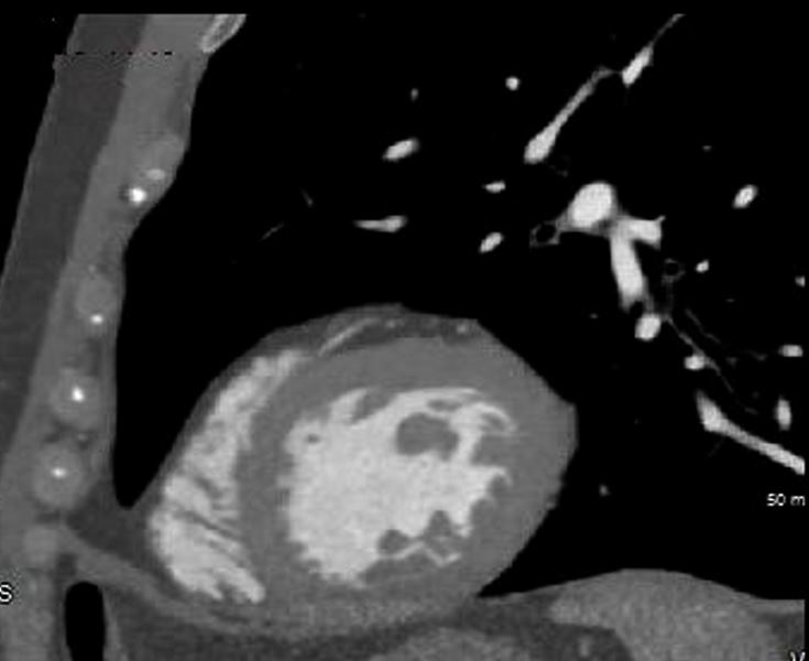
Septum Apex Lateral Walls in Coronal Projection and the Papillary Muscles

key words right atrium heart cardiac RA tricuspid valve TV left atrium LA MV mitral valve RV right ventricle anterolateral papillary muscle interventricular septum left ventricle LV CT scan
Ashley Davidoff MD
34780
Septum Inferior Posterior and Anterior Walls in Sagittal Projection
Ashley Davidoff MD
Anterior, Apical Inferior and Basal Portions in a Sagittal 3D Reconstruction
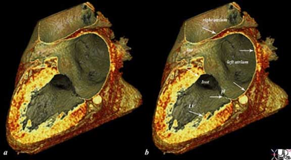
The reconstructed CT scan is in the same plane as the autopsy specimen above, and shows a distended left ventricle (LV), left atrium (LA) and right atrium (RA). The thin endocardium (white arrows) throughout the heart is one continuous sheet of tissue connected across the whole circulatory system and it is more fibrous in nature. The thin white layer is seen in the left atrium, left atrial appendage (LAA) over the posterior leaflet of the mitral valve (pl) in the left ventricle (LV) and in the right atrium. The surface of the left ventricle, left ventricular outflow tract (LVOT) and right atrium are also lined by the endothelium.All images courtesy: Ashley Davidoff, M.D.
47824c02.8s
Short Axis in 3D at the Base Showing Mitral to Aortic Fibrous Continuity, Septum, Inferior and Lateral Walls

Thickness
The sagittally reconstructed CT study of the heart demonstrates the normal LV thickness, chamber size, mitral valve with continuity to the aortic valve. The smaller of the two hollow tubes to the left of the aorta represents the left atrial appendage and the larger more superior structure represents the main pulmonary artery
.
Davidoff MD 47823 copyright 2009 all rights reserved
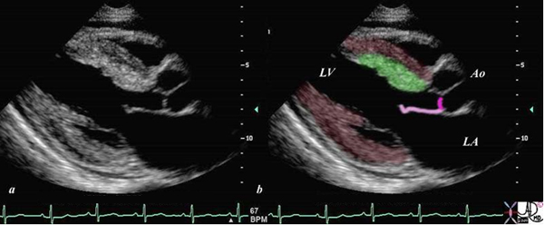
Echo Showing Fibrous Continuity of the Anterior Leaflet of the Mitral Valve to the Non Coronary Cusp of the Aortic Valve
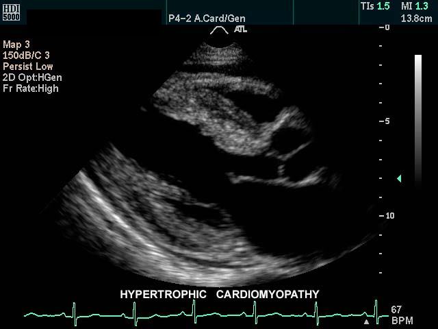
This gray scale echo of the heart shows the left ventricle, anterior (light pink) and posterior leaflets of the mitral valve, the aortic valve (dark pink), and the base of the aorta. There is a focal thickening of the ventricular septum (green) in the left ventricular outflow tract just proximal to the aortic valve. The region is also slightly more echogenic than the remaining myocardium (maroon). This case demonstrates a case of asymmetric septal hypertrophy or muscular subaortic stenosis.
Courtesy Philips Medical Systems 33134 33134c04.8s
tags IHSS, ASH
Ashley DAvidoff MD
heart-anatomy-P-039