
Principles Imaging
Copyright 2009
Introduction
The golden years of medical imaging are now. We have come from the basic x-ray examination in 1895 via advanced X-ray in the form of angiography in the 1950’s, ultrasound and nuclear imaging in the 1960’s, CT in the 1970’s and MRI in the 1980’s . The field has blossomed over the last decade and we cannot imagine contemporary medial practice without any of these modalities, and wonder about the next horizon which will likely be related to nanotechnology.
In all instances the principles of imaging remain constant.
|
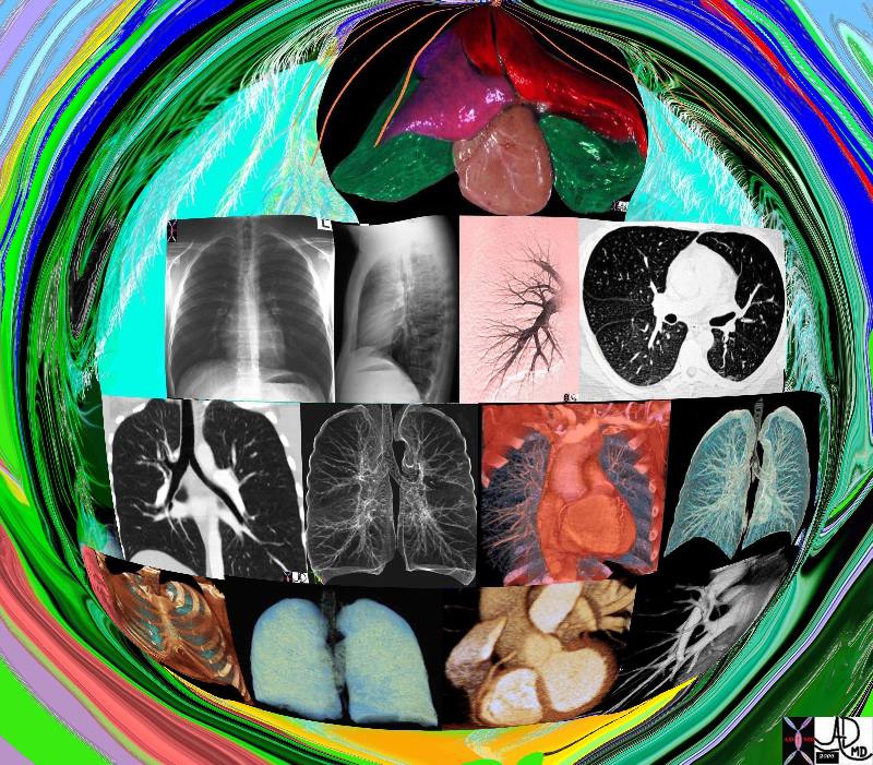
Imaging the Cardiopulmonary Team |
| 42444b17.800 lung chest pulmonary artery bronchus bronchi trachea heart normal anatomy Ddavidoff art Davidoff MD |
Aim
The true reflection of structure and function is the goal of imaging.
For every action there is an equal and opposite reaction
In imaging we interrogate structure by thrusting certain forces (X-rays, US waves, radiofrequency waves) toward the object to be imaged and the object reacts to the force in specific and characteristic ways reflecting its nature and makeup. Thus if we use X-rays as our force then some of the X-rays we thrust upon the structure to be interrogated are totally reflected, some partially reflected and some pass through without reflection. This interaction allows us to define the elements discussed including size, shape, position, character, connections and relations. If we can repeat the interrogation of the same structure at a different time then we can evaluate how it changes with time and circumstance and hence have a window into how the structure functions. In the case of the heart the relevant changes occur every millisecond, and cycle every second, while in the case of the ovary and the endometrium, the relevant physiological events cycle through a month.
Imaging and structure
As we have outlined in the introduction to the common vein, structure is defined by very consistent and specific elements.
All structures have;
As the science of imaging advances, the ability of the tools to reflect the true nature of the structure becomes truer and truer to reality.
Imaging and function
While structure has been within the realm of traditional radiological imaging, the techniques that have allowed us to image how structure changes with time have given us a window into the functional aspects of the body. Thus echocardiography reflects motion of cardiac muscle and cardiac valves, doppler imaging reflects flow in arteries and veins, perfusion imaging reflects, as implied, perfusion of tissue with contrast agents, and diffusion imaging reflecting Brownian motion.
Nuclear imaging with radioisotopes has been a longstanding technique for functional imaging. For many years it not only enabled us to study contractile abilities of muscle as well as blood flow but also enabled us to identify how certain metabolites were handled by the body. The most recent innovation of this technique called PET scanning reflects for example how sugar is handled by the tissues. Diseases such as inflammation and cancer have heightened metabolic needs and hence heightened need and use of glucose. This metabolic need is reflected in the imaging technique and metabolic activity and hence function is reflected.
Structure and function in biology are packaged in one gift, and the one without the other is impossible. Dividingthese two and studying them separately is the huma way of attempting to understand them.
Indications
Medical imaging is indicated when a person is suspected of suffering from a disorder and accurate diagnosis eludes the usual preliminary investigations that include taking a clinical history, examining the patient and running a battery of preliminary blood tests.
Contraindications
Absolute contraindications to imaging are rare. There are a few relative contraindications to certain imaging techniques under specific physiological or clinical situations. For example the use of X-rays during the period of featal organogenesis is relatively contraindicated. There are alternatives in this situation. For example ultrasound would be considered quite safe during this period.
MRI is contraindicated in patients with pacemakes, certain metallic implants, or metallic foreign bodies that have inadvertantly been implanted while working.
Intravenous contrast is relatively contraindicated in patients with severe contrast allergy, or patients with renal failure. Claustrophobia is a real problem for some patients and this is also a relative contraindication. However in general, medical imaging is quite safe with relatively few contraindications.Patients who are allergic to intravenous contrast would
Advantages
The advantages of medical imaging is to enable more accurate diagnosis of structural and or functional abnormality in order to provide optimal therapy
Disadvantages
Medical imaging is stressful, sometimes uncomfortable, costly, and has risks of radiation exposure.
Method
Patient Preparation
It is imperative for all the studies that the patient is well prepared both physically and psychologically. For a gallbladder using ultrasound, it is imperative that the patient is in the fasting state since ingestion of food causes the gallbladder to contract and limits visiualization and therefore evaluation of the gallbladder.
A nuclear medicine study is a two part examination. There is first the injection of the radioisotope, and then image acquisition. These may be hours or even a day apart. The time the patient has to deote to the study should be well explained. The patient also has to lie quietly for 20-40 minutes during acquisition. The patient has to be able to do this otherwise the study may be unreadable.
The patient has also to be prepared psychologically for the exam and to know what to expect. The MRI study requires that the patient lie still in the gantry, while being subjected to a battery of uncomfortable noisesA patient arricing for a nucleatr medicine study should know how long the study takes and also that they are required to lie still.
Equipment
Not all equipment is equal. The difference between an 8slice MDCT scanner and a 64MDCT scanner is enormous. Similalrly there are strengths and limitataions based on technology and manufacurer.
Techniques
Plain film radiography requires accurate positioning of the patient, accurate positioning of the equipment, and appropriate utilization of Kv and mA. Ultrasound evaluation is operator and patient dependent. Operator’s skill and experience varies significantly and hence the results obtained will vary. CT is less operator dependant and overall good studies are usual even when the patient is large. MRI is dependant on protocols provided by the radiologist since there are so many sequences that each bring out different characteristics of the disease process. The patient has to be still in the scanner and any movement will degrade the image significantly.
Results
(1) Component parts.
(2) A size or a dimension.
(3) Shape.
4) Position in space.
(5) Character, i.e. when a structure interacts with its environment there are characteristic ways in which it will behave. For example, elastic behavior, liquid behavior, hard soft, green blue.
(6) Change with time
7) All biological structures have relationships with other structures, and are connected to other structures and environments. The connections take the form of conduits that bring products in to the structure, conduits that take products out of the structure (arteries, veins, capillaries and lymphatics), or connect to the environment directly in intimate contact.
Complications
In general diagnostic imaging has few complications. The application of diagnostic imaging to guided procedures do have inherent procedure related complications but those are not considered complications of the imaging devices but rather of the procedure.
Contrast Differences

How Many Squares in the Centre of the Image? |
| 72772 contrast difference gray scale imaging pinciples of X-ray Davidoff Art Courtesy Ashley Davidoff MD 72772 72773 72774 72775 72775b01 72776 72777 |

Gray Scale |
| 72773 contrast difference gray scale imaging pinciples of X-ray Davidoff Art Courtesy Ashley Davidoff MD 72772 72773 72774 72775 72775b01 72776 72777 |
Air
•
Air floats – high point
Air is black
Fluid
•
Fluid drops – low point
Fluid is white