
- 50yo
-
- history of stage IVB SCC cervix
- mets to lungs, liver, bones, vagina
- L3 pathologic fracture,
- recent L2 compression fracture,
- prior DVT
- recent
- significant progression of metastatic disease
- history of stage IVB SCC cervix
-
MRI 5 years ago
-
- Enhancing mass with restricted diffusion that measures approximately 4.5 cm centered in right aspect of the cervix with involvement of the right upper vagina and right parametrial extension.No lymphadenopathy.
-
MRI Uterus Cervix and Vagina
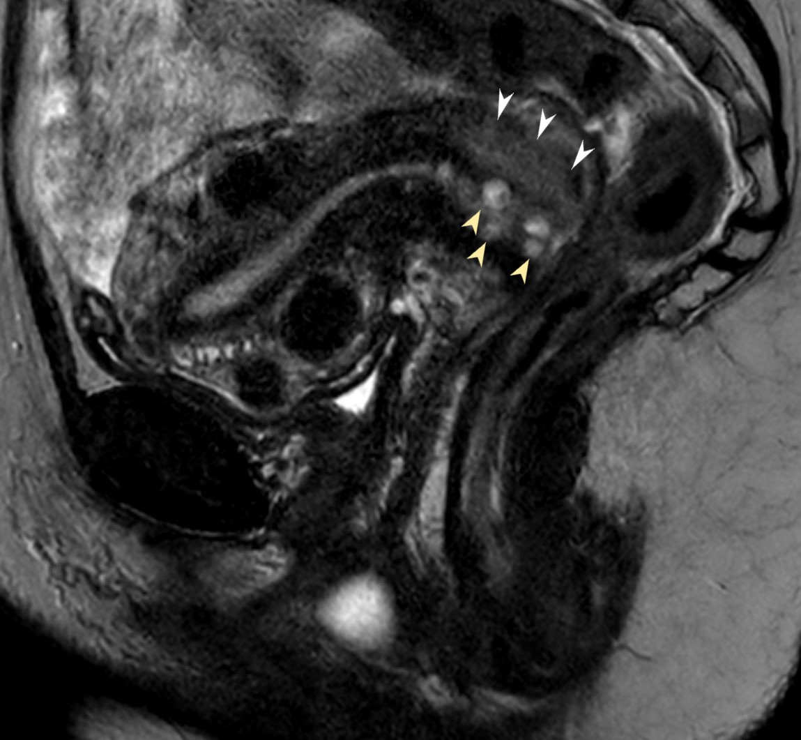
MRI in the sagittal plane using T2 weighted imaging shows a soft tissue mass on the superior aspect of the cervix (white arrowheads). Incidental note is made of multiple Nabothian cysts (yellow arrowheads)
Ashley Davidoff MD TheCommonVein.net 135659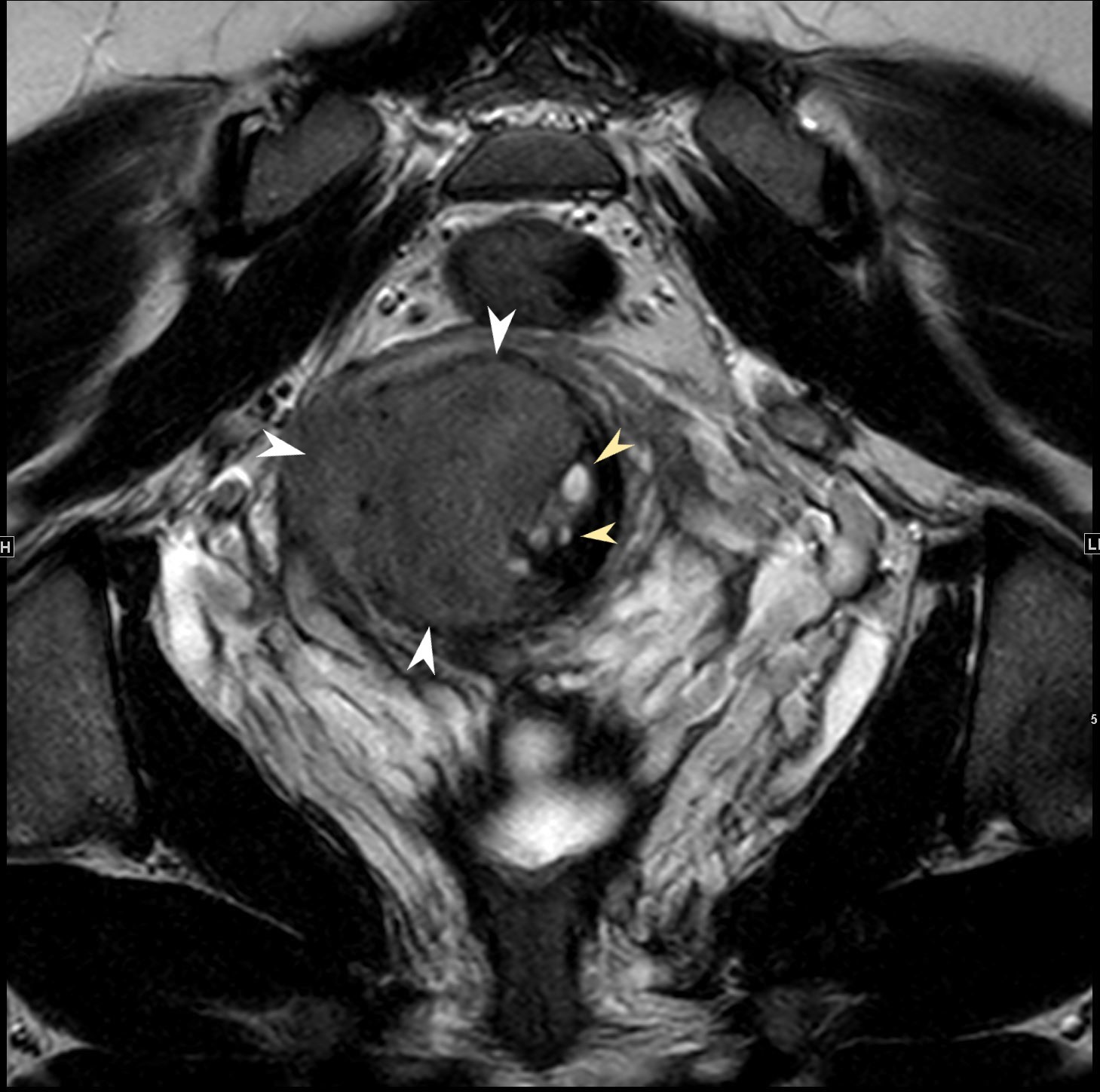
MRI Cervix
MRI in the coronal plane through the cervix using T2 weighted imaging shows a large soft tissue mass on the lateral aspect of the cervix (white arrowheads). Incidental note is made of multiple Nabothian cysts (yellow arrowheads)
Ashley Davidoff MD TheCommonVein.net 135660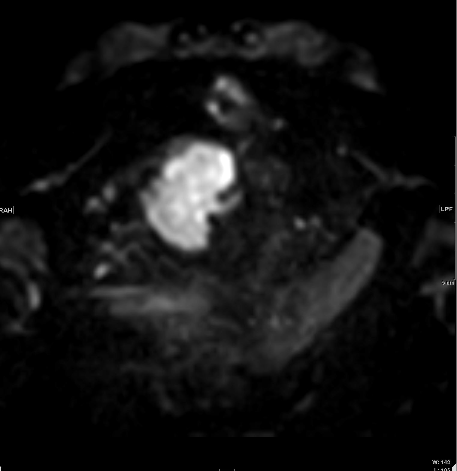
PET Scan Cervical Mass
PET scan in the coronal plane through the cervix shows a large hyperintense mass on of the cervix
Ashley Davidoff MD TheCommonVein.net 135661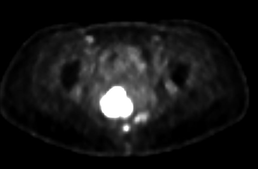
PET Scan Cervical Mass
PET scan in the axial plane through the cervix shows a large hyperintense mass on the lateral aspect of the cervix
Ashley Davidoff MD TheCommonVein.net 135662
5years Later After Chemo Radiation she Presents with
Intermittently Tachycardic to 110s
Imaging Shows Bulky Mediastinal Adenopathy
Encasing
Pulmonary Arteries, Main Stem Bronchi Pulmonary veins and Obstruction of the Esophagus
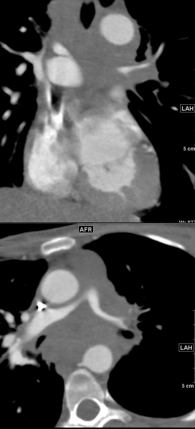
Encasement of the Main Stem Bronchi 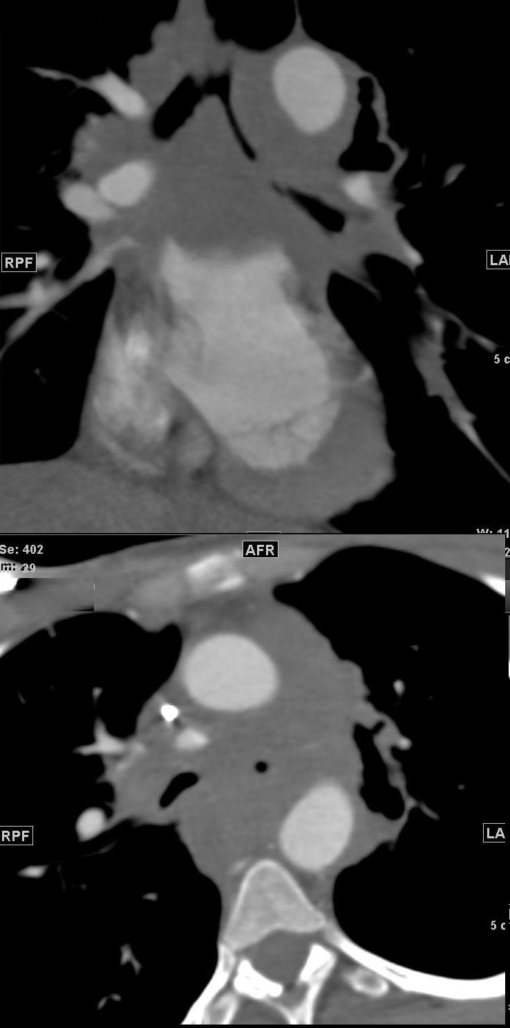
CT of the Mediastinum Encasement of the Bronchi
50-year-old female with a history of metastatic carcinoma of the cervix. CT in the coronal plane through the mediastinum shows bulky nodal metastases that encase the left main stem bronchus in the upper image, and extends to encase both bronchi better seen in axial projection in the lower image.
Ashley Davidoff MD TheCommonVein.net 135663
Encasement of the Pulmonary Arteries
50-year-old female with a history of metastatic carcinoma of the cervix. CT in the coronal plane through the mediastinum shows bulky nodal metastases that encase the left pulmonary artery in the upper image, and extends to encase both left and right main pulmonary arteries, better seen in axial projection in the lower image.
Ashley Davidoff MD TheCommonVein.net 135664
Encasement of the Pulmonary Veins and Right Lung Congestion
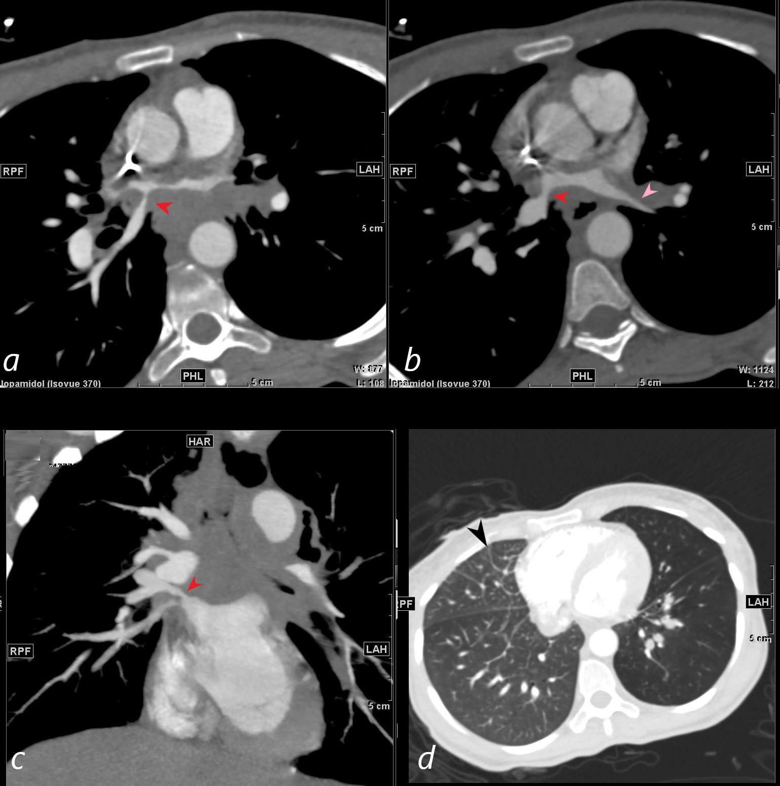
50-year-old female with a history of metastatic carcinoma of the cervix. CT through the upper left atrium shows focal narrowing of the right upper pulmonary vein (a b and c red arrowheads) with upstream dilation and mass effect on the posterior wall of the left atrium caused by bulky nodal metastases. There is diffuse encasement of the left upper pulmonary vein (b pink arrowhead). Image d through the lungs show asymmetric congestion of the right lung with prominent Kerley B lines (black arrowhead), likely from the more severe narrowing of the right sided pulmonary veins centrally.
Ashley Davidoff MD TheCommonVein.net 135666
Obstruction of the Esophagus
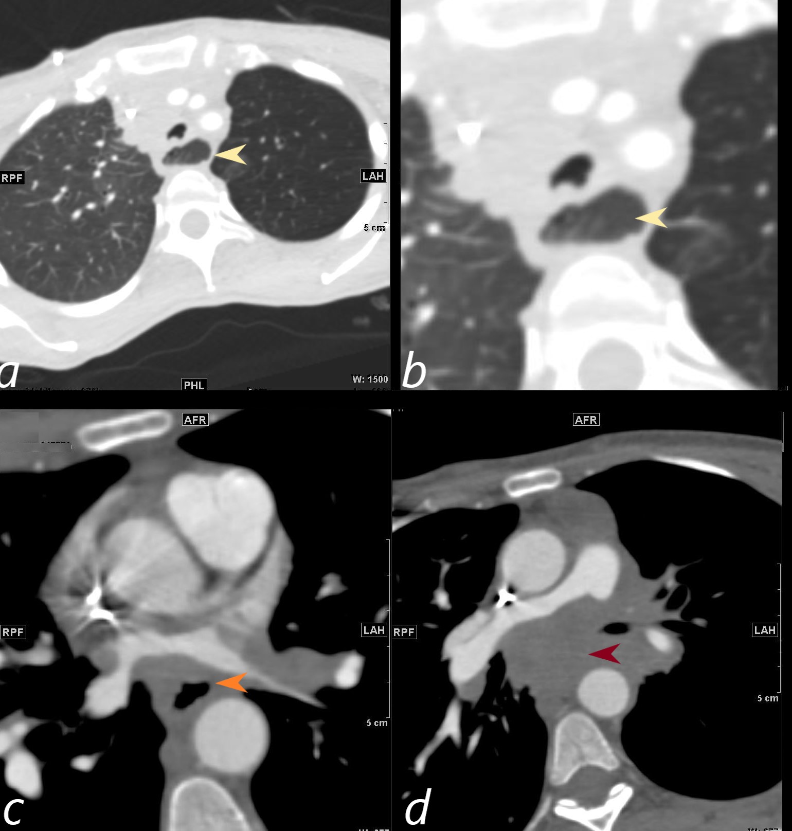
50-year-old female with a history of metastatic carcinoma of the cervix. CT through the chest shows dilation of the proximal esophagus that contains fluid and secretions (a, magnified in b, light yellow arrowheads) becoming narrower due to the mass effect (c orange arrowheads) and finally effaced and obliterated (d maroon arrowhead).
Ashley Davidoff MD TheCommonVein.net 135668