
Left Ventricular Aneurysm
Copyright 2008
- Definition
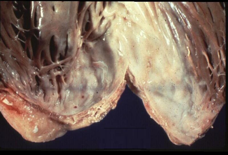
- well delineated, thin, scarred, or fibrotic wall
- devoid of muscle or containing necrotic muscle,
- Causes of Left Ventricular Aneurysm and Pseudoaneurysm
-
- Post MI –
- caused by total occlusion of the left anterior descending coronary artery and the absence of collateralization
- RCA inferoposterior basal wall
- Circumflex lateral wall
- patients with multivessel disease,
- aneurysms are uncommon due to
- extensive collateralization
- patients with multivessel disease,
- hypertrophic cardiomyopathy and
- Chagas disease,
- Post MI –
-
- Result
- Functional
- akinetic (without movement) or
- dyskinetic
- CHF
- Structural
- Size 1-8 cms
- Shape
- Round
- Position
- apex and anterior wall are 70 to 85 %
- inferoposterior basal wall RCA
- lateral LVA secondary to left circumflex occlusion is exceedingly rare
- Character
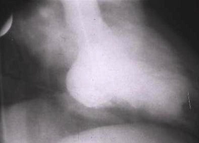
Study from 1960
Ashley Davidoff
THE COMMONVEIN.net
- The wall of the aneurysm typically consists o
-
- white fibrous scar and is very thin.
- organized clot in over 50 percent of cases
-
- Causes of Left Ventricular Aneurysm and Pseudoaneurysm
-
- Post MI –
- hypertrophic cardiomyopathy and
- Chagas disease,
- Complications
- CHF
- ventricular arrhythmias
- arrhythmias
- Rupture – rare
-
Ashley Davidoff
THECOMMONVEIN.net

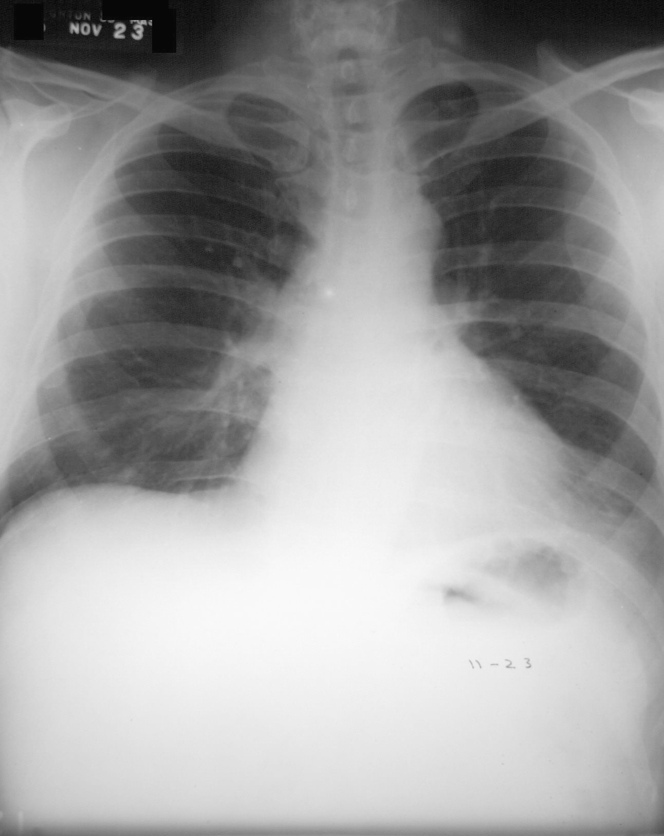
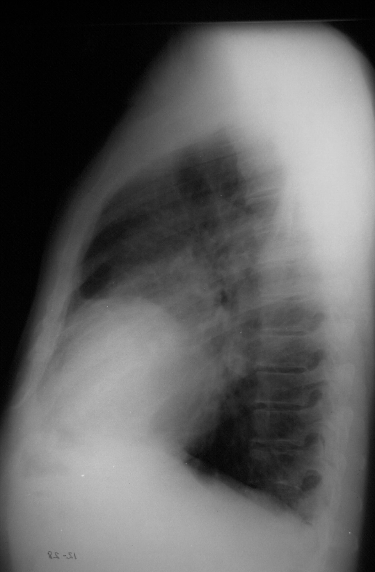
Dr Lloyd Hawes

Courtesy Dr Lloyd Hawes
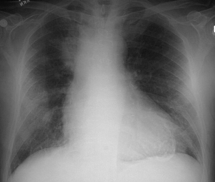
TheCommonVein.net
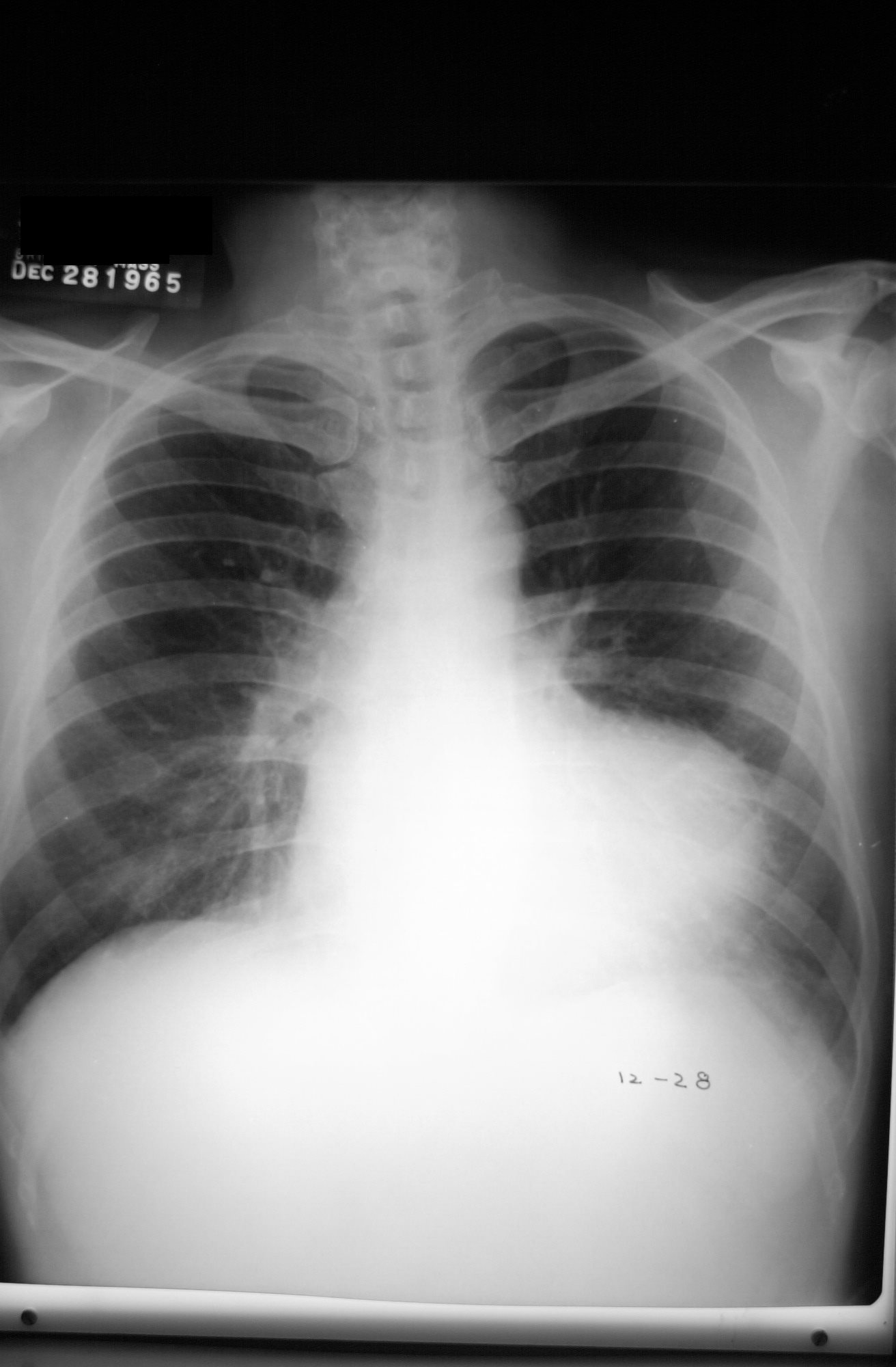
Courtesy Dr Lloyd Hawes
TheCommonVein.net
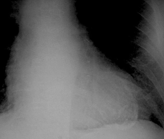
1 month later
Courtesy Dr Lloyd Hawes
TheCommonVein.net
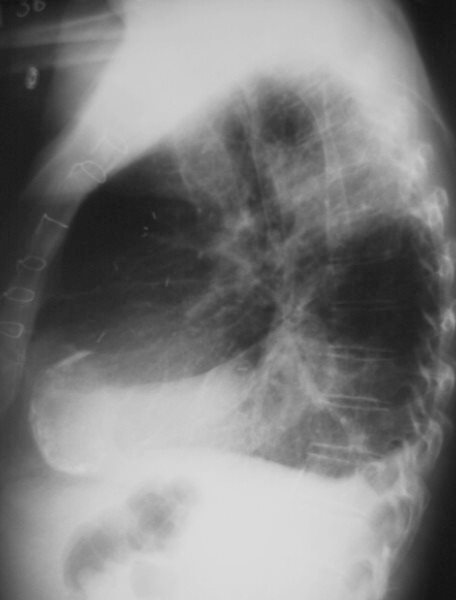
Courtesy Dr Lloyd Hawes
TheCommonVein.net
Ashley Davidoff
TheCommonVein.net.
Ashley Davidoff
TheCommonVein.net.
Ashley Davidoff
TheCommonVein.net.

The CommonVein.net
The CommonVein.net
The CommonVein.net
The CommonVein.net
The CommonVein.net
The CommonVein.net
The CommonVein.net
TheCommonVein.net
TheCommonVein.net
TheCommonVein.net
TheCommonVein.net

RCA distribution
TheCommonVein.net
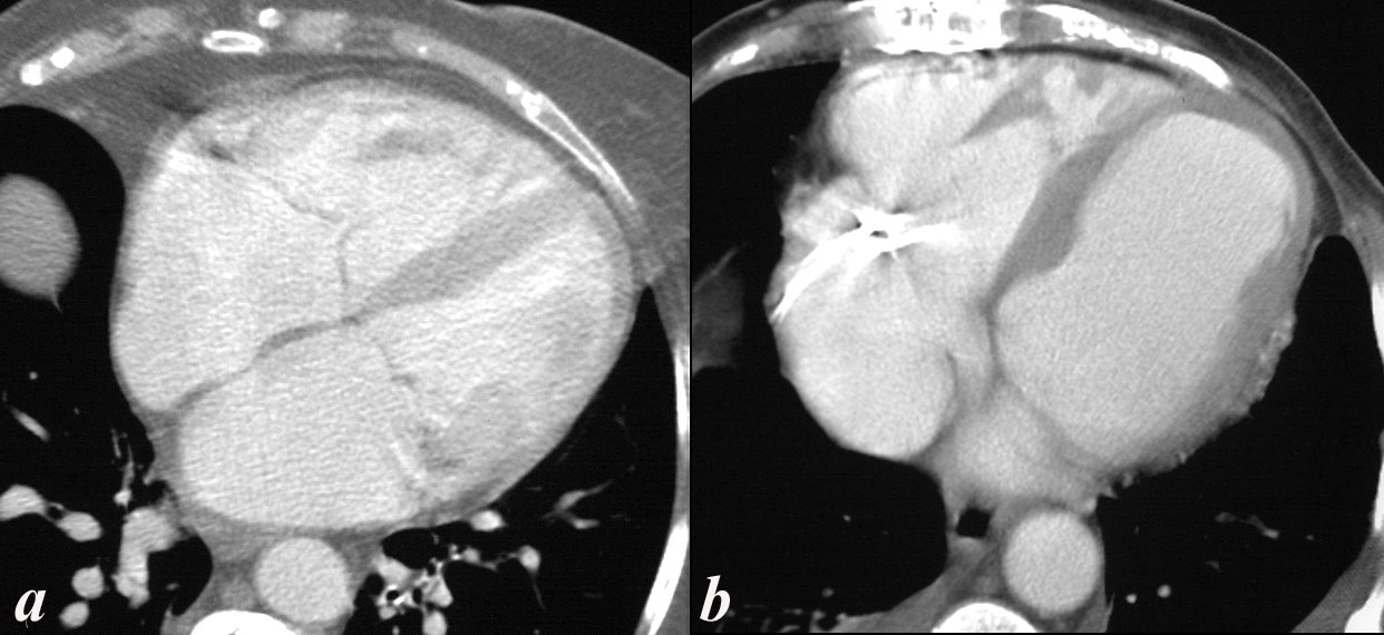
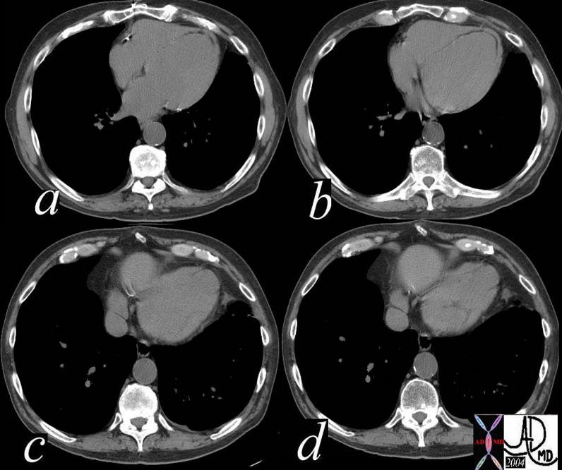
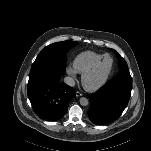
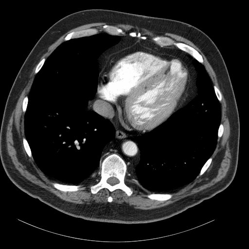
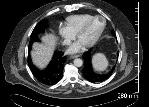
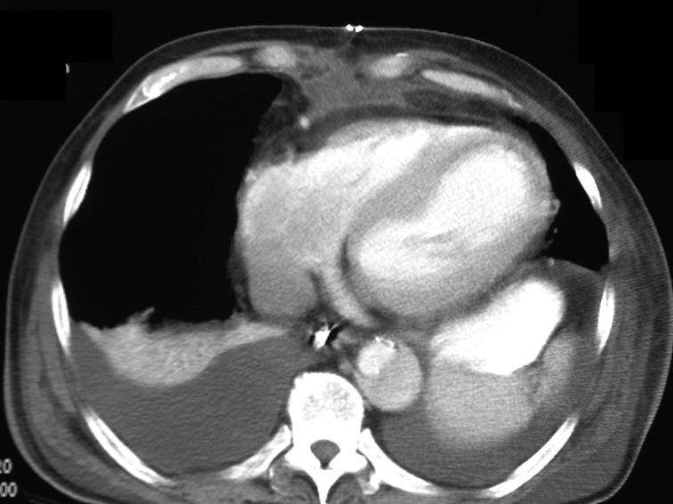
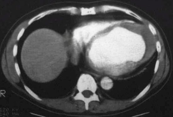
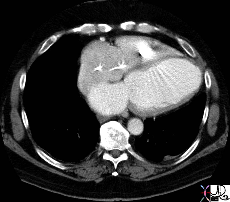
Axial CT through the left ventricle (LV) shows rounding of the apex and thinning of the overlying myocardium. This finding is consistent with an apical LV aneurysm most likely caused by LAD disease.
Note the fat dysplasia in the apical myocardium which confirms the prior infarct.
Ashley Davidoff MD
0040 B heart 57 M aneurysm
Note also a dissection in the descending aorta
Ashley Davidoff
THECOMMONVEIN.net

Ashley Davidoff
THECOMMONVEIN.net
|
01213 |
| Courtesy Ashley Davidoff MD 01213 esophagus + fx dilated + fx fluid filled + dx achalasia of the cardia +cardiac heart LV apex left ventricle calcified calcification aneurysm thrombus RS pleura pleural calcification asbestos related disease imaging radiology CTscan |
|
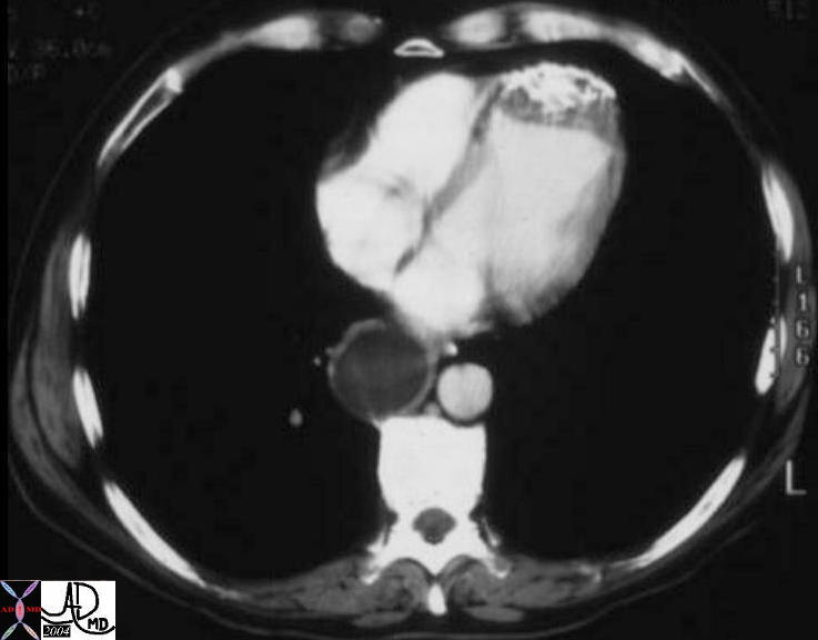
30472 |
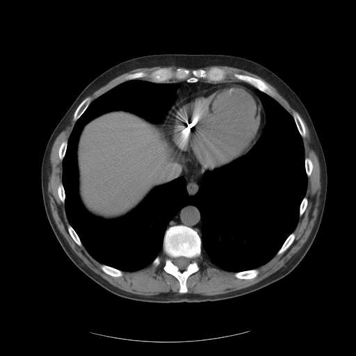
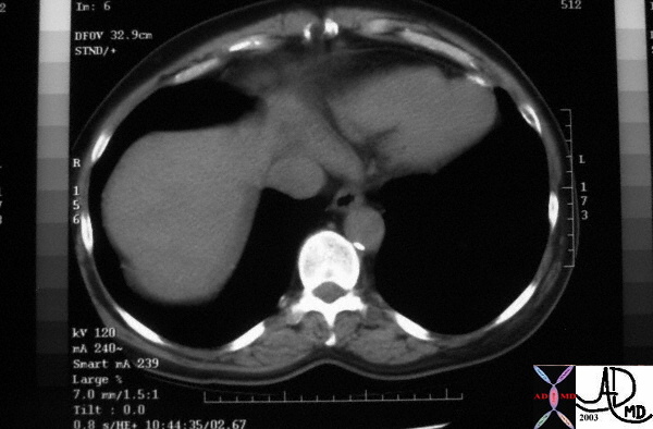
| This cross sectional CT image of the heart shows calcified apex of the left ventricle associated with thrombus, characteristic of an LV aneurysm. The cause is almost certainly secondary to coronary artery disease and ischemic heart disease with secondary myocardial infarction. Courtesy Ashley Davidoff MD. 30472 code heart LV apex IHD CAD aneurysm calcification calcified MI cardiac imaging radiology CTscan |
|
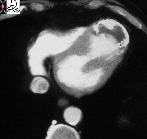
30473 |
| This cross sectional CT image of the heart shows calcified apex of the left ventricle associated with thrombus, characteristic of an LV aneurysm. The cause is almost certainly secondary to coronary artery disease and ischemic heart disease with secondary myocardial infarction. Courtesy Ashley Davidoff MD. 30473 code heart LV apex IHD CAD aneurysm calcification calcified MI cardiac imaging radiology CTscan |
|
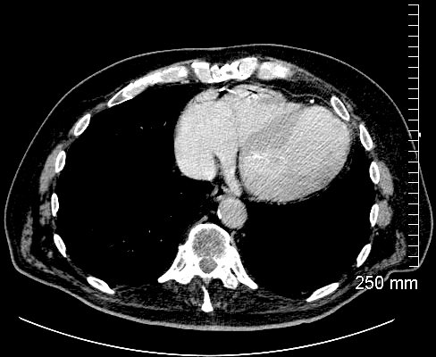
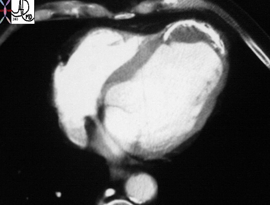
32074 |
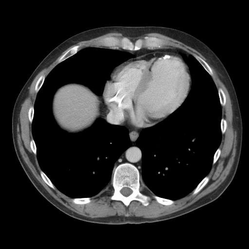
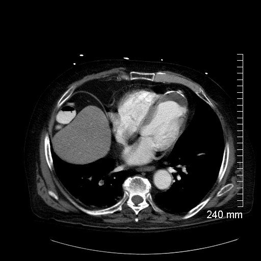
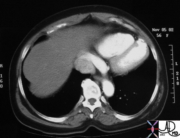
| This cross sectional CT image of the heart shows the apex of the left ventricle associated with thrombus, characteristic of an LV aneurysm. The cause is almost certainly secondary to coronary artery disease and ischemic heart disease with secondary myocardial infarction. Courtesy of Ashley Davidoff M.D. 32074 code heart LV apex aneurysm thrombus aneurysm IHD CAD MI large cardiac imaging radiology CTscan |
|
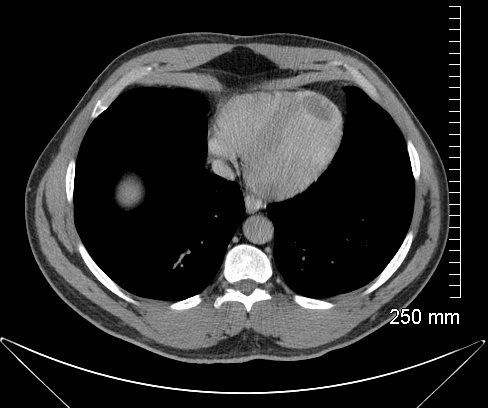
33511 |
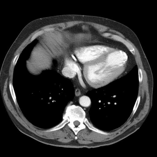
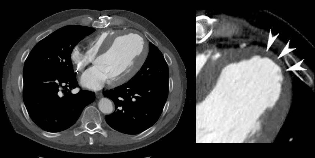
| This transverse CT scan through the apex of the left ventricle shows a focal calcification at the apex. The diagnosis evolves into a focal pseudoaneurysm of the LV apex. Courtesy Ashley Davidoff MD 33511 code heart LV apex calcified pseudoaneurysm inflammation immune rheumatic heart disease RHD cardiac imaging radiology CTscan |
|
33157 |
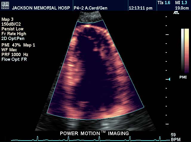
| This power motion imaging echo of the heart showing a 4 chamber view, and demonstrating the left ventricle. The apex of the LV is rounded suggesting a LV aneurysm. Courtesy Philips Medical Systems 33157 code cardiac heart echo LV aneurysm dyskinetic segment apex imaging cardiac echo |
42394.800 |
| 42394.800 heart cardiac LV left ventricle platypus shape fx dilated fx enlarged dyskinetic segment CTscan Davidoff MD |
Pseudoaneurym at the base
References and Links
TCV