
Fossa Ovalis
Copyright 2008
Definition
The fossa ovalis is an oval depression on the septal wall of the atrium, that occupies the middle portion.
Structurally it consists of the central fibrous flap valve called the septum primum that is surrounded by a ring of muscle called the septum secundum. The central septum primum has oval shaped defect superiorly that is called the foramen ovale reminiscent of its shape.
Functionally the septum primum enables blood to flow from the right atrium to the left atrium in fetal life. Post natally the pressure in the right atrium rises and causes the flap valve to close, though the valve remains potentially open in about 30% of adults. The superior portion of the septum secundum is called the superior limbic band but also is known as the crista dividens, because it was thought to direct (dividens =divide) the flow of SVC blood in the fetus, into the right atrium and away fronm the foramen ovale.
Diseases of the fossa ovalis are almost exclusively related to diseases of the septum primum, and include ASD of the secundum type and patent foramen ovale.
Diagnostic elements relate to these diseases and include the clinical evaluation for an atrial septal defect (systolic murmur, split fixed P2, right ventricular enlargment, and pulmonary hypertension later in life) EKG showing enlarged RV, and echocardiography that can document defects in the fossa ovalis.
Treatment is usually surgical.
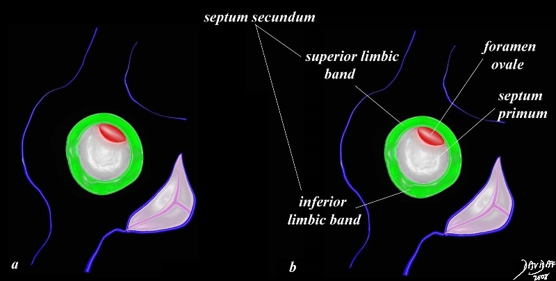
The fossa ovalis is a central structure of the atrial septum that consists of a ring of muscle (green) surrounding a membrane called the septum primum, that has a crescentic defect at the 1 oclock position (red) called the foramen ovale. The superior portion of the muscle is called the superior limbic band, and the inferior aspect is called ythe inferior limbic band.
01656c04.8s
Ashley Davidoff MD
Davidoff
TheCommonVein.net
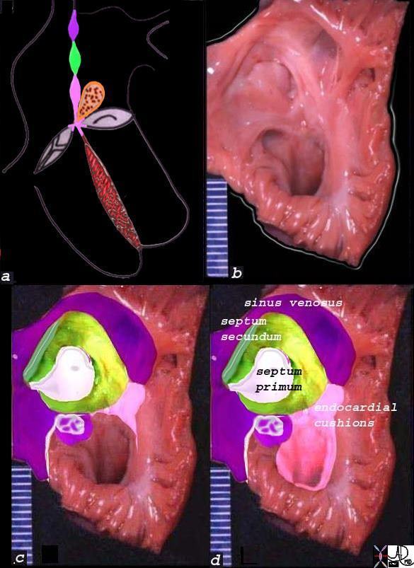
The diagram shows the three portions of the interatrial septum in (a) with the upper portion (purple) deriving from sinus venosus tissue, the middle (grreen) the mesodermal tissue, and the lower (pink) the endocardial cushion tissue. Image b, is an anatomical specimen that is overlaid in reference colors in c, and labelled in d. The middle of the atrial septum is a fibrous membrane called the septum primum, and it is surrounded a rim of muscular tissue called the septum secundum.
The fossa ovalis is the middle portion of the interatrial septum and consists of the central portion called the septum primum (white) and the surrounding muscle called the septum secundum (green).
Ashley Davidoff MD
01653c11b05a04 TheCommonVein.net
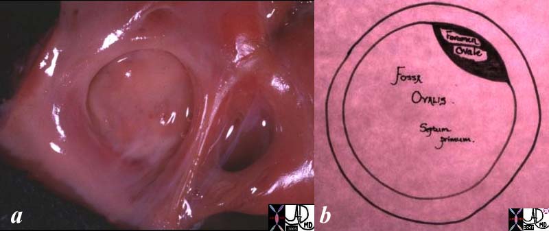
The fossa ovalis is the depressed ring like structure surrounded by a rim of muscle seen in the first image, consisting of the septum primum and foramen ovale seen in the second diagram.
01671c.8s
Ashley Davidoff MD
TheCommonVein.net