
The Nerve Supply
Ganesh Athappan MD and Ashley Davidoff MD
Conduction System of the heart
Introduction
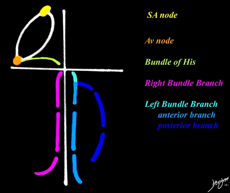
The systematic contraction and relaxation of the cardiac chambers to facilitate cardiac functioning is coordinated by the conduction system of the heart. The nervous system of the heart is structurally composed of specialized muscle fibers that generate and transmit cardiac impulses. The major components of controlling sites include the sinoatrial node (SA node), atrioventricular node (AV node), the bundle of His, the left and right bundle branches, and subendocardial branches of Purkinje fibers.
Structural Considerations:
The conduction system, like the vascular system, is built around the scaffolding of the heart.
Relationship of the Conduction System to the Scaffolding |
|
The conduction system is shown around the scaffolding of the heart. The SA node (yellow) is situated high in the right atrium near the SVC, connects with the A-V node (orange) through unnamed specialized conduction cells to which is situated at the entrance of the IVC into the right atrium. The bundle of His (green) transmits the impulse along the AV groove and the conduction system then penetrates ventricular septum at the crux, to divide into the right (pink) and left (blue) branches. The left branch has an anterior (light blue) and a posterior division (royal blue). Courtesy Ashley DAvidoff Davidoff art copyright 2009 all rights reserved 07915b03.8s |
Sinoatrial node:
The Sinoatrial node (also called the sinus node SA node ,SAN) is the impulse generator, and is located in the RA close to the opening of the SVC. It is made of specialized cells that have the ability to self-initiate an electrical impulse. It is composed of a small cluster of fusiform cells located in the sulcus terminalis on the subepicardial surface at the SVC RA junction. In contrast, the A-V node is situated in a subendocardial position. It is characterized by an intrinsic automaticity that ‘fires’ at a rate that is faster than other pacemakers and hence by this virtue becomes the impulse generator of the heart. Once the SA node initiates an electrical impulse, the resulting electrical wave moves its way across the right and left atria and to the AV node.

Position of the SA Node |
|
The right atriogram in the A-P projection reveals contrast in the SVC, IVC and right atrium (RA). The SA node (yellow) lies at the junction of the SVC with the right atrium 06635c.81s Courtesy Ashley Davidoff copyright 2009 all rights reserved |

Subepicardial position in the Sulcus Terminalis |
|
The sulcus terminalis (blue line) is a junctional line seen on the external surface of the right atrium running between the SVC and IVC . The SA node lies on the subepicardial surface, on the sulcus terminalis, at the junction of the SVC with the RA (yellow sphere) on the sulcus terminalis (yellow sphere) Courtesy Ashley DAvidoff MD copyright 2009 all rights reserved 06628cc03.8s |

Sulcus terminalis on the Inside of the Heart |
|
The anatomical specimen exhibits a right atrium that has been opened showing the SVC superiorly (superior royal blue ring), the inferior vena cava (blue ring inferiorly) with its associated Eustachian valve (purple), the entrance of the coronary sinus (small light blue ring) and the annulus of the tricuspid valve (teal ring). The SA node (yellow) lies close to the entrance of the SVC on the sulcus terminalis Courtesy Ashley DAvidoff MD copyright 2009 all rights reserved 06630b03bc01b03.8s |
The blood supply of the SA node is usually from the right coronary artery (60%) and sometimes from the left circumflex. (40%)
Arterial Supply of the SA node |
|
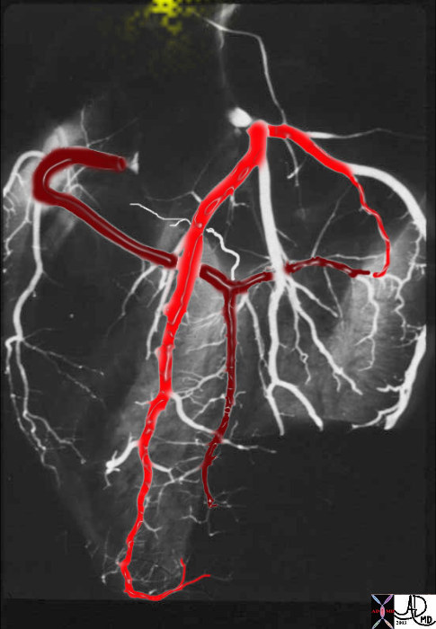
This post mortem angiogram in the LAO projection shows normal right (RCA) and left (LCA) coronary arteries. The LCA is overlaid in bright red, and the RCA in maroon. The S-A node (yellow) is seen in the superior aspect of the RA. 33805SA Courtesy Ashley Davidoff MD. Copyright 2009 |
AV node
The atrioventricular node is an area of specialized tissue between the atria and the ventricles of the heart and is positioned at the posteroinferior region of the atrial septum, occupying the apex of the triangle of Koch.
The base of triangle of Koch is formed by the coronary sinus, with each of the other limbs formed by the Eustachian valve posteriorly and a portion of the tricuspid annulus anteriorly where the septal leaflet of the tricuspid valve attaches.

Triangle of Koch |
|
The specimen exhibits a right atrium that has been opened showing the SVC superiorly (royal blue ring) the inferior vena cava (blue ring inferiorly) with its associated Eustachian valve (purple), the entrance of the coronary sinus (small light blue ring) and the annulus of the tricuspid valve (teal ring). The triangle of Koch is shown with its base formed by the coronary sinus, a posterior limb formed by the Eustachian valve (ligament of Todaro) and the anterior limb formed by the annulus of the tricuspid valve. The AV node lies within this triangle. The SA node (yellow) lies close to the entrance of the SVC 06630b03bc03.8s Courtesy Ashley Davidoff MD copyright 2009 all rights reserved |

Triangle of Koch – Magnified |
|
The right atrium has been opened in this normal heart revealing the atrial septum and the fossa ovales and the annulus of the tricuspid valve The purpose is to demonstrate the triangle of Koch, which has its base in the coronary sinus, one limb along the Eustachian valve (ligament of Todaro) and the other along the tricuspid annulus. The A-V node (orange sphere) lies within the triangle. 08452bb01c04b08b02b.8s Courtesy Ashley Davidoff MD copyright 2009 all rights reserved |
The AV node measures about 3-5mms in diameter, and is situated in the subendocardial region. In contrast the SA node is situated in a subepicardial position. The AV node delays impulses for about .12seconds before allowing impulses through to the His-Purkinje conduction system, which in turn spreads impulses to the ventricular walls. This delay ensures that the atria have ejected their blood into the ventricles before the ventricles contract.
The blood supply to the AV node is via the AV nodal artery that arises from the posterior crux of the coronary circulation and is a branch of the right system in almost 85% of the time.

Blood Supply to the AV node |
|
The post mortem angiogram in the LAO projection shows the main branches of the left, non dominant circulation in bright red and the right dominant coronary system in maroon. The SA node and pathways(overlaid in yellow) connect with the AV node (orange) . The AV nodal artery in this instance, and in almost 85% of hearts arises from the right system at the posterior crux of the heart (white arrow). 33805SAAV01b03.8s Davidoff MD copyright 2009 all rights reserved copyright 2009 |
The AV node dissipates its signals to the ventricles via the His bundle. At the junction of the membranous and muscular ventricular septum the His Bundle penetrates the septum and divides into right and left bundles or branches.

Bundle of His |
|
The post mortem angiogram in the LAO projection shows the left non dominant circulation in bright red and the dominant right coronary artery in maroon. The SA node and pathways(yellow) to the AV node (orange) is shown from which the Bundle of His (green) proceeds to the interventricular system near the crux of the heart 33805SAAV05.8s Davidoff MD copyright 2009 all rights reserved copyright 2009 |
Right and Left Bundles
The right and left bundle branches proceed on either side of the ventricular septum deep to the endocardium and ramify into subendocardial branches which form the Purkinje fibers. The left bundle in turn branches into left anterior and posterior fascicles.
The left fascicle runs down the left side of the ventricular septum, covered by endocardium. In the lower part of the left ventricle, the bundle breaks up into numerous strands which end in the papillary muscles and in the ventricular muscle. These strands between the endocardium and the cardiac muscle are called Purkinje fibers.
Right and Left Bundles |
|
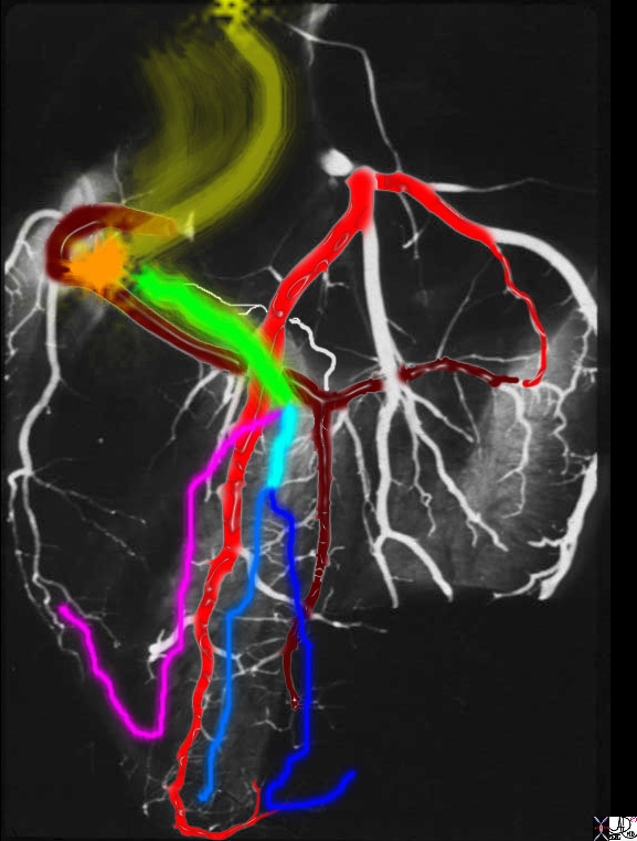
The post mortem angiogram in the LAO projection shows the left non dominant circulation in bright red and the dominant right coronary artery in maroon. The SA node and pathways (yellow) to the AV node (orange) is shown from which the Bundle of His (green) proceeds to the interventricular system near the crux of the heart. The conduction system penetrates the membranous septum and divides into the right bundle branch (RBB = pink) and the left bundle branch (LBB = teal blue) which rapidly divides into the anterior (light blue) and posterior divisions (royal blue) . The RBB and anterior division of the left bundle are supplied by the septal perforators of the LAD and the posterior bundle (royal blue ) is supplied by the septal perforators of the PDA 33805SAAV07.8s Davidoff MD copyright 2009 all rights reserved copyright 2009 |

The Left Bundle Branch and its Divisions |
|
The post mortem specimen has been overlaid to demonstrate the left bundle branch (yellow) of the conducting system as it travels within the left side of ventricular septum. The left bundle enters below and between the right coronary cusp and the non coronary cusp through the membranous septum. The right coronay cusp houses the right coronary ostium (rco) that is represented as a rounded red circle, and the left coronary cusp houses the left coronary ostium (lco). The non coronary cusp is situated between the two coronary cusps. The anterior leaflet of the mitral valve is in fibrous continuity with the left coronary cusp and non coronary cuspp Once the left bundle enters the left side of the septum,it divides into an anterior and a posterior division. Davidoff art copyright 2009 07970ec03.8s 07970ec03.8s |
The muscle of the heart has an intrinsic ability to conduct nerve stimuli as evidenced by the modification of these cells to form the conduction system. In addition the muscle is organized and is characterized by a syncitial network so the cells are in open contact with each other. The myocytes have an “open door” structure meaning there are breaks in their cell membrane that allows for the optimal conduction and coordination of the nervous impulse enabling coordination of the muscular impulse.

Cardiac Syncitium |
|
This is an artistic rendition of the cardiac myocytes. The cardiac myocytes are organized in a syncitium so that the cells have an “open door” structure with each other. The conduction pathway is shown as white arrows (b) and some of the “doors” between the cells are overlaid with green rings to indicate the break in the cell membrane that allows for the free passage of conductivity of the nerve impulse enabling coordinated muscular activity that is required in cardiac contraction. 32906b05c04.8s Davidoff art Courtsey Ashley Davidoff MD Copyright 2009 All rights reserved |
Autonomic Nerve Supply
The nerves that supply the heart are derived from the cardiac plexus, which are formed partly from the parasympathetic vagi and partly from the sympathetic trunks. They are freely distributed both on the surface and in the substance of the heart, the separate nerve filaments being furnished with small ganglia. Vagus fibers are inhibitory, or depressor, nerves. Sympathetic nerves are accelerator nerves.
The sympathetic system causes an increase in cardiac rate, and contraction force, and causes the coronary arteries to become dilated by acting on the SA node, AV node, and directly on the atria and ventricles.
The parasympathetic system slows the cardiac rate and decreases the force of contraction by acting on the SA node, AV node, atria, but has only sparse innervation of the ventricles.
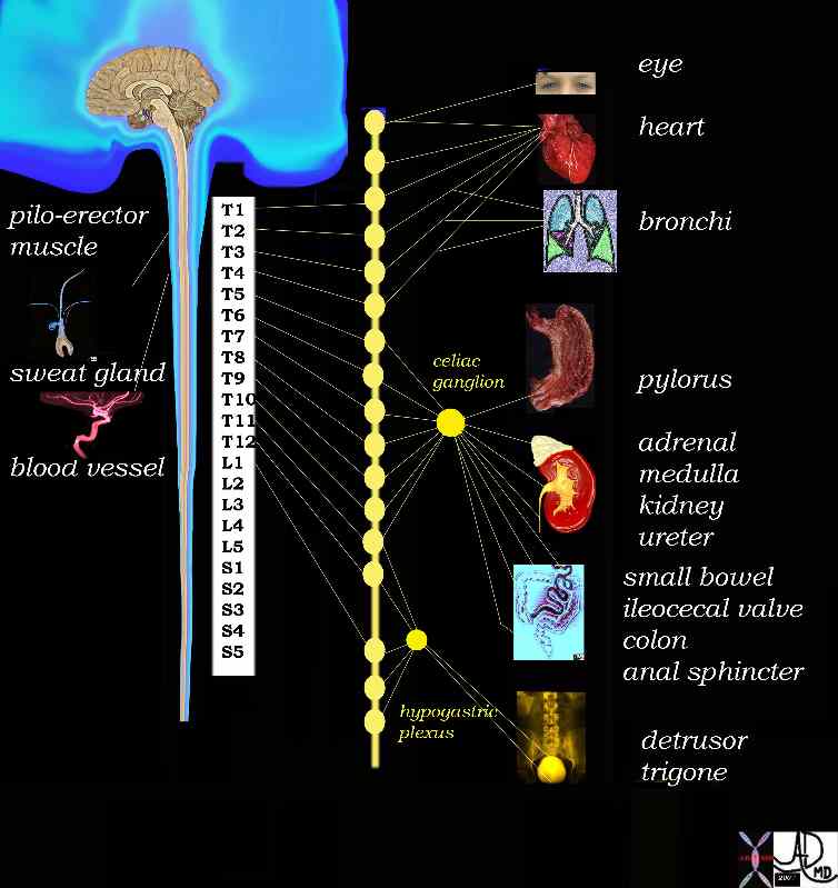
Sympathetic Efferent System |
|
The sympathetic fibers the lower cervical region, as well as from T1- T 4 contribute to the sympathetic tone of the heart . 14798d03.800 Davidoff Art Davidoff MD Copyright 2009 |
|
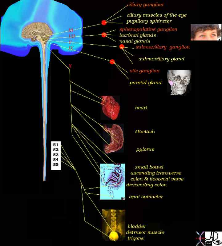
Parasympathetic Efferents |
|
The vagus nerves both right and left contribute to the sympathetic control of the heart. They arise as the 10th cranial nerves and represent the efferent system of the parasympathetic system to the heart and other organs. 14798d06 copyright 2009 Davidoff Art Davidoff MD |
Clinical Case
|
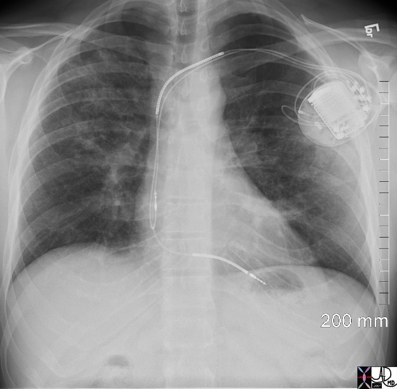
Normal CXR and one with Pacemaker needed for Complete Heart Block Secondary to Involvement of the Conduction System of the Heart by Sarcoidosis |
|
CXR is from a 34 year old man with sarcoidosis. The sarcoidosis also affected his myocardium and AV node causing complete heart block for which he required an emergency life saving pacemaker. The electrode in the right atrium is coiled on itself to enable close contact with the right atrial wall, and the second is in the right ventricular apex and controls the ventricular rate. 49740b03.800 Copyright 2009 Davidoff MD |
Blood supply to the conduction system is provided by both the right and left systems, but dominantly by the right which supplies the SA node in 60%, the AV node by the right in 90%, and the posterior fascicle of the left bundle in 90%. The left coronary artery supplies the SA node in 40%, AV node and bindle of His in 10%, the right bundle and the anterior fascicle of the left bundle in almost all cases.