
Rheumatic Heart Disease
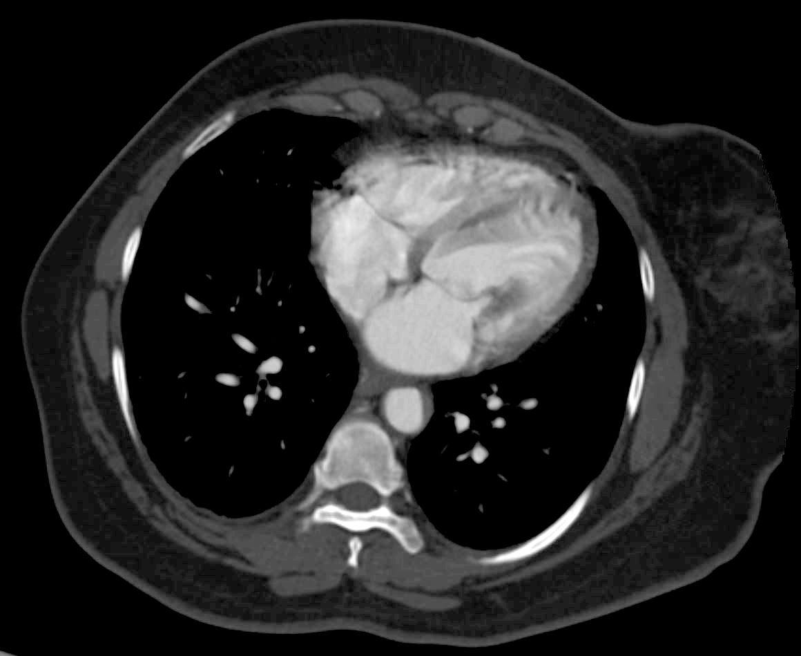
Axial CT through the heart shows a papillary muscle close to the insertion of the valve indicating shortened chordae. This is a finding associated with rheumatic heart disease. Echo showed evidence of both mitral stenosis and mitral regurgitation.
Ashley Davidoff MD TheCommonVein.net 135548
Axial CT through the heart shows a normal papillary muscle and normal chordae.
Ashley Davidoff MD TheCommonVein.net 135505

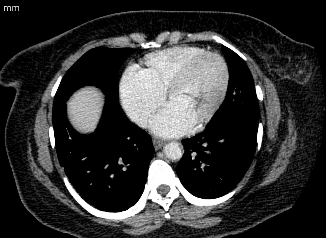
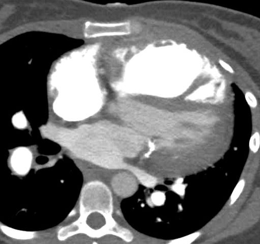
87 year old male with findings consustent with mitral stenosis , characterised by calcified mitral valve, enlarged left atrium, normal sized left ventricle,with enlarged right atrium, right ventricle and main pulmonary artery.
Pure mitral stenosis is most commonly caused by rheumatic heart disease
Ashley Davidoff MD
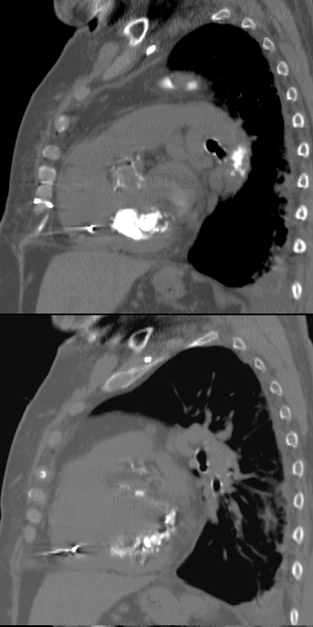
87 year old male with findings consistent with mitral stenosis , characterised by calcified mitral valve, enlarged left atrium, normal sized left ventricle,with enlarged right atrium, right ventricle and main pulmonary artery.
Pure mitral stenosis is most commonly caused by rheumatic heart disease
Ashley Davidoff MD
50 year old female with a history of rheumatic mitral stenosis
CT with contrast in the axial plane shows a mildly thickened mitral valve in systole
Courtesy Ashley Davidoff MD TheCommonVein.net 136519
Libman Sacks Vegetation
SLE and PULMONARY HYPERTENSION without ILD
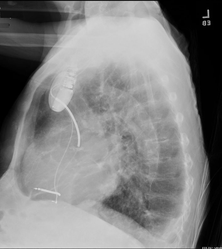
27-year-old female presents with dyspnea and a past history of SLE, Raynaud’s disease, and Lupus nephritis.
Chest X-ray shows cardiomegaly with right ventricular configuration on the PA and an enlarged main and probably left pulmonary and an enlarged descending RPA. The lateral confirms the enlarged RV and raises the possibility of LV enlargement.
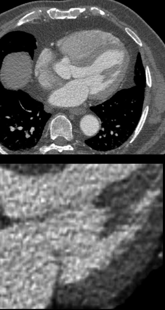
The CT scan confirms an enlarged MPA, RPA, RA and RV, and shows calcification on the posterior leaflet of the mitral valve consistent with Libman Sacks vegetation. There is mild ground glass opacity at the lung bases but no sign of ILD.
A current non contrast abdominal CT shows a pericardial effusion and normal sized kidneys
Ashley Davidoff MD
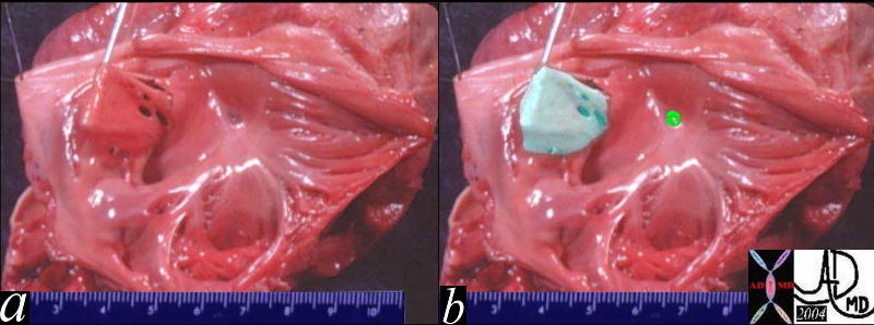
Vegetation

The four chambered echocardiogram of the heart shows an echogenic mass representing a mitral valve vegetation characteristic of endocarditis.
key words
heart cardiac MV mass SBE bacterial endocarditis vegetation infection imaging cardiac echo overlay
Courtesy of Philips Medical Systems, Ultrasound, and modified by Ashley Davidoff M.D 32121
Annulus Calcification
82-year-old female with end stage renal failure, diabetes mellitus, CAD, s/p CABG x2 and AVR, severe mitral annular calcification, extending to the septum, s/p pacemaker, with associated mild mitral stenosis and significant mitral regurgitation. Severe aortic stenosis warranted AVR.
Sagittal reconstruction of the CT of the chest shows severe mitral annular calcification extending into the ventricular cavity and region of the septum. Note sparing of the anterior leaflet of the mitral valve.
Ashley Davidoff MD
Congenital Mitral Atresia
Congenital AVC Canal
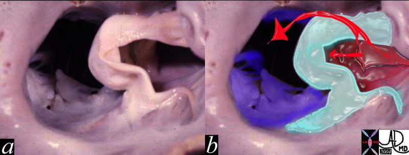
Complete Endocardial Cushion Defect
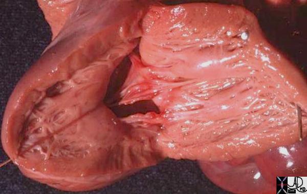
The pathology specimen shows a complete endocardial cushion defect with a cleft mitral valve and a VSD. The atrial chambers can be seen through the cleft of the mitral valve and a ASD primum was present as well
Key words
heart cardiac LV left ventricle IVS interventricular septum mitral valve endocardial cushions complete AV canal defect cleft mitral valve VSD endocardial cushion defect congenital heart disease gross pathology Courtesy Ashley Davidoff MD 01863b01
Congenital Mitral Stenosis
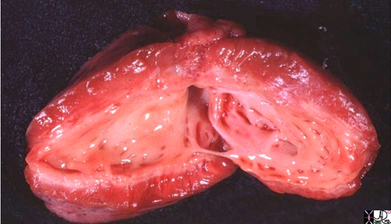
The gross pathology specimen is a case of hypoplastic left heart with aortic stenosis and mitral stenosis, revealing a thickened endocardium with left ventricular hypertrophy and deformed and thickened mitral valve and papillary muscles. The thickened endocardium is caused by the high pressures generated in the LV causing subendocardial ischemia and resulting in a condition called endocardial fibroelastosis (EFE). Images courtesy of: Ashley Davidoff, M.D. 08058.8s