
heart0067-low res

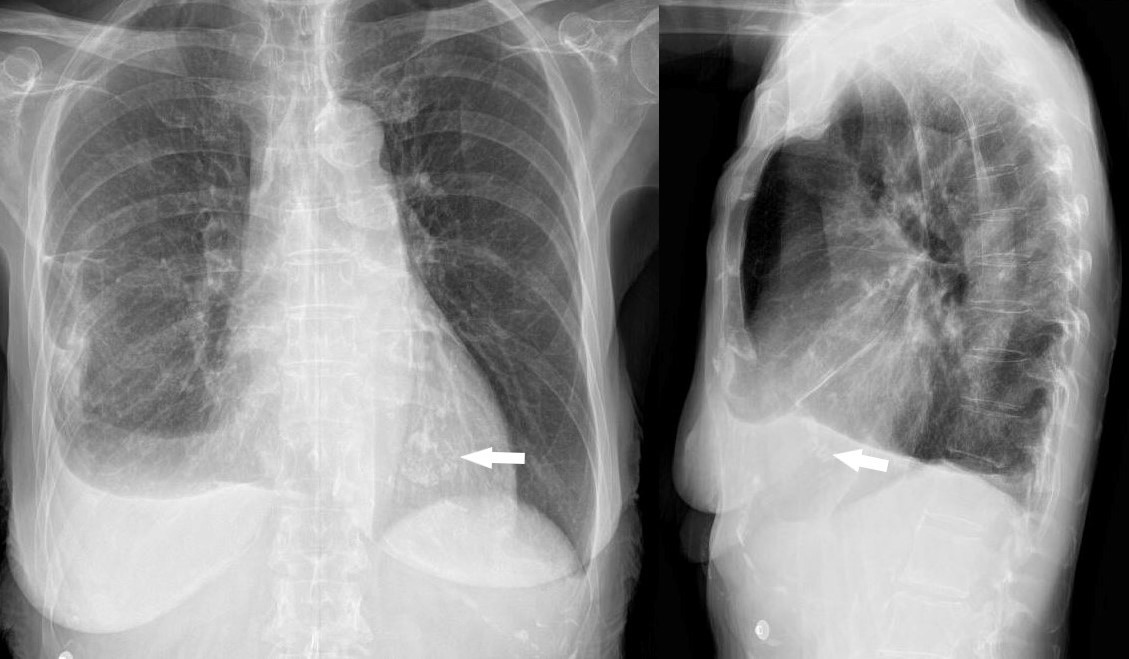
The patient is a 73 year old female with apical hypertrophic disease. She has a history of diastolic heart failure, pulmonary hypertension, CREST, esophageal stricture, COPD and chronic renal failure
heart0068b-low res
Ashley Davidoff MD
The patient is a 73-year-old female with apical hypertrophic disease. She has a history of diastolic heart failure, pulmonary hypertension, CREST, esophageal stricture, COPD and chronic renal failure
heart0069b02 Ashley Davidoff MD
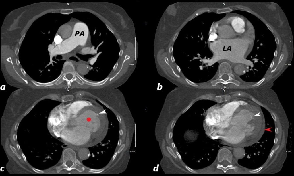
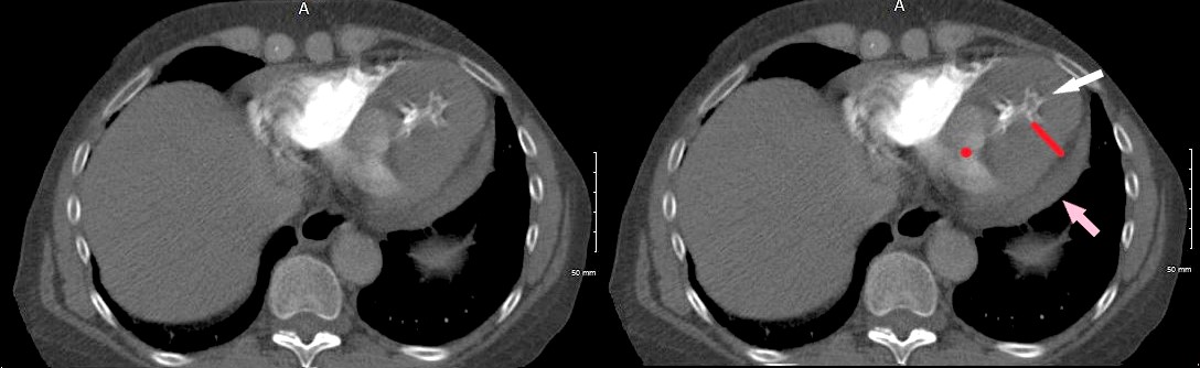
The CT axial images shows an enlarged pulmonary artery (PA) indicating pulmonary hypertension, and I an enlarged left atrium (LAE, b) , a small left ventricular (LV) cavity (red circle , c) calcified foci in the LV (c and d white arrows, and a small pericardial effusion (red arrow d)
The patient is a 73-year-old female with apical hypertrophic disease. She has a history of diastolic heart failure, pulmonary hypertension, CREST, esophageal stricture, COPD and chronic renal failure
Ashley Davidoff MD
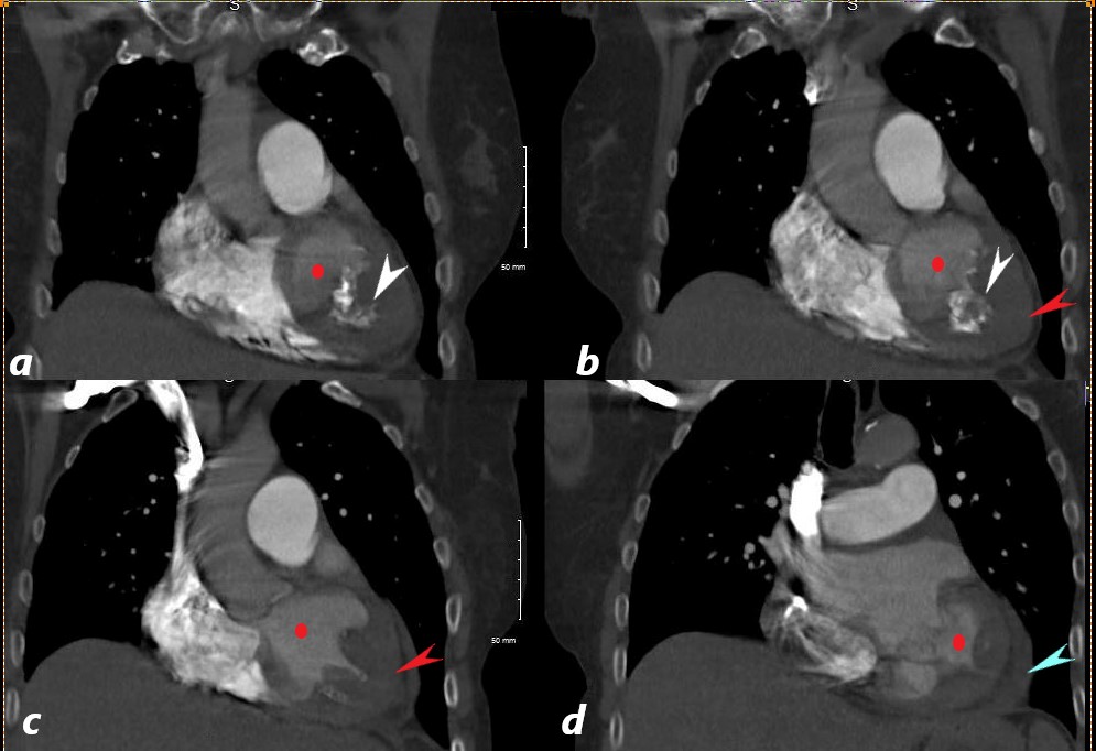
The CT coronally reconstructed images show a small left ventricular (LV) cavity (red circle ,a,b,c,d) intracavitary calcification (white arrow a,b), apical LV hypertrophy (LVH, red arrows b, and c) and a small pericardial effusion (blue arrow, d)
The patient is a 73-year-old female with apical hypertrophic disease. She has a history of diastolic heart failure, pulmonary hypertension, CREST, esophageal stricture, COPD and chronic renal failure
Ashley Davidoff MD
References and Links
- TCV