Tubes of the Cardiovascular System
Copyright 2009
Introduction
The heart is positioned at the center of all the organs in the body and it is understandably considered one its vital organs. The circulation is dependant on the vascular system consisting of a variety of tubes that enable delivery of blood to the organs, the perfusion of the cells, and then the collection of blood and metabolic products for the return journey to the heart. The principles governing the function and flow in the tubes is universal , but each of the tubes is designed with a specific purpose with unique structure and therefore unique function.
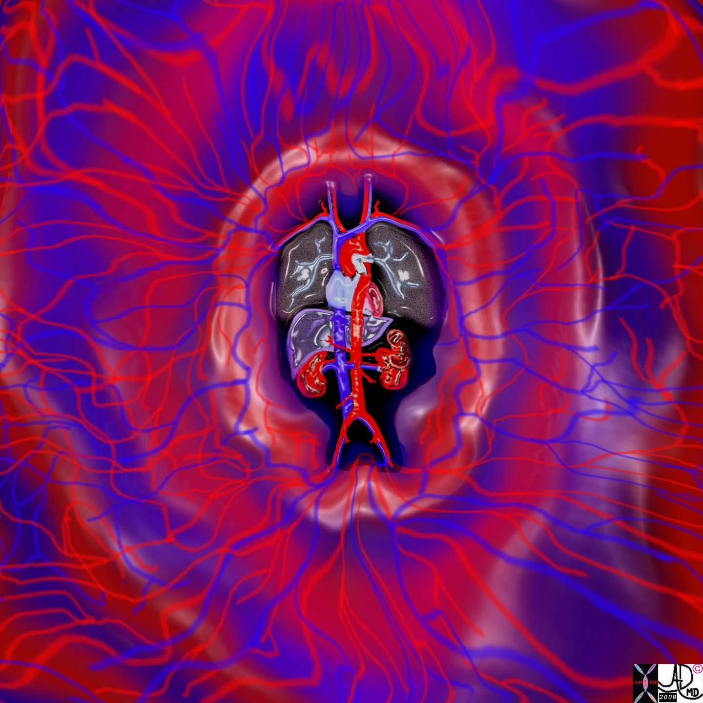
This image provides an overall view of the heart and its component tubes consisting of arteries, capillaries and veins.
32059.41ks heart cardiac circulation arteries veins capillaries pulsation pulsatile flow normal anatomy physiology
Ashley Davidoff MD art copyright 2008 chronicle high resolution
The function of transport is the domain of the tubes of the body, which have to obey the universal principles of flow within a tube. If you understand flow in tubes, you will be able to extend the principes to a large portion of of the way the body works in health and the consequences when the transport systems fail in disease.Tubes and Transport
Flow will only occur when a pressure difference exists between the two sides of the tubes. When we breathe in, the intercostal muscles contract and expand the chest laterally and the diaphragm moves down, increasing the volume of the chest cavity in a craniocaudad dimension. As a result the alveoli expand, causing a negative pressure in the alveolar side of the tubes. The pressure in the atmosphere is higher and thus air will flow from a high pressure system to a low pressure system – i.e. from the atmosphere to the lungs. During expiration, the elastic nature of the chest, with the relaxation of intercostal muscles and diaphragm, result in a return of the chest to its preinspiratory position. The chest therefore is in a contracted size, causing a relative increase of pressure in the alveoli when compared to the atmosphere, and air will flow from the alveoli to the atmosphere. Inspiration is therefore an active process requiring energy and muscle contraction, and expiration is a passive process of elastic recoil.
In the cardiovascular system the heart acts as a pump to increase the pressure on the arterial side so that flow will occur from the high pressure to the low pressure. In the gastrointestinal tract the peristalsis of smooth muscle creates the upstream high pressure so that products of ingestion and digestion can be brought to the factories of absorption and eventually for evacuation.
Velocity of flow relates to factors such as tubular diameter, resistance, friction, and pressure differences, as well as the nature of the medium being transported. Velocity of flow will also depend on whether the flow is laminar or turbulent. If it is laminar, it will be governed by Poiseuille’s law, which states that velocity of flow is directly proportional to the driving pressure.
Poiseuille’s law – for laminar flow
Volume flow rate = pressure difference
resistance
= P1-P2
R
= pressure difference X radius4
8/pi viscosity X length
As velocity increases however, flow tends to become turbulent and Poiseuille’s law does not apply. Flow tends to be laminar when the tubes are small, and turbulent when the tubes are large or when there are irregularities in the walls. Thus flow in the larger parts of the airways tends to be turbulent and in the smaller tubes it is laminar. Under resting conditions, laminar flow exists from the medium-sized bronchi onward down to the bronchioles. During exercise, the air flow is accelerated, and laminar flow may be confined only to the very small airways. Laminar flow is quiet while turbulent flow is noisy. Thus in the chest examination, the sound of wheezing or the presence of murmurs indicates turbulent flow which is abnormal. The audible “huffing and puffing” that occurs with exercise results from greater forcefulness of the muscles in an attempt to get more air in and out, and the turbulence becomes audible even without the stethoscope. Similarly audible wheezing in asthmatic patients reflects narrowing of the smaller airways.
The function of transport is the domain of the tubes of the body, which have to obey the universal principles of flow within a tube. If you understand flow in tubes, you will be able to extend the principes to a large portion of of the way the body works in health and the consequences when the transport systems fail in disease.
Laminar Flow
|
|

Turbulent Flow
|
|