
Buzz
- Pancarditis
- pericardium, pericarditis 25% most common
- myocardium, myocarditis is rare caused by vasculitis
- myocardial infarction 9X increase
- endocardium – Libman-Sacks 10% mitral and tricuspid valve
- Cardiac complications in about 50% and major cause of death
Pericarditis
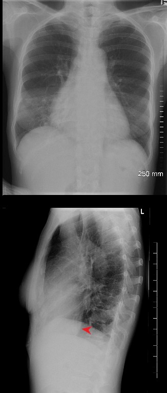
2 views of the chest of a 54 year old female with SLE and Sjogren’s syndrome show a slightly enlarged heart in the PA projection and an unusually prominent and rotund IVC (red arrowhead)
Ashley Davidoff MD
key words
pericarditis, pericardial effusion, SLE
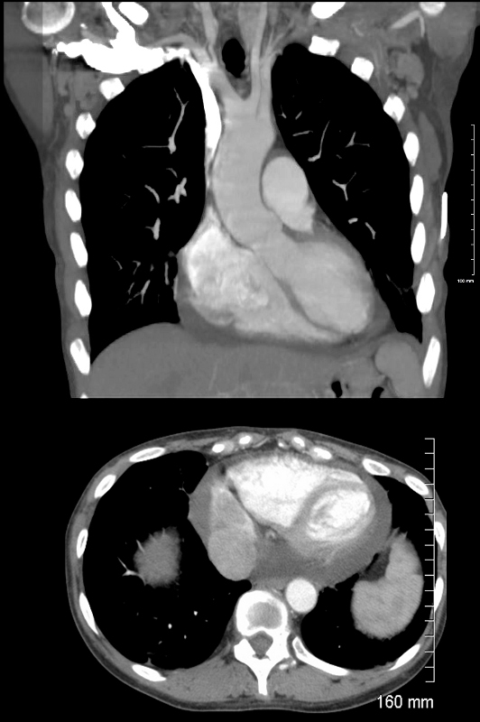
Coronal (above) and axial images CT showing the Rv and LV of a 54 year old female with SLE and Sjogren’s syndrome. A small pericardial effusion is noted accounting for the cardiomegaly on CXR.
Ashley Davidoff MD
key words SLE, pericarditis, pericardial effusion
Myocarditis
occurs in nearly 10% of SLE patients and serves as a prominent contributor of the heart failure (HF) and poor prognosis 8, 29 and is caused by vasculitis with with microvascular coronary dysfunction. SLE myocarditis is an immune complex-mediated process.
The LGE is characterized by patchy and small areas in the midwall and subepicardial myocardium in both the both acute and chronic phases4

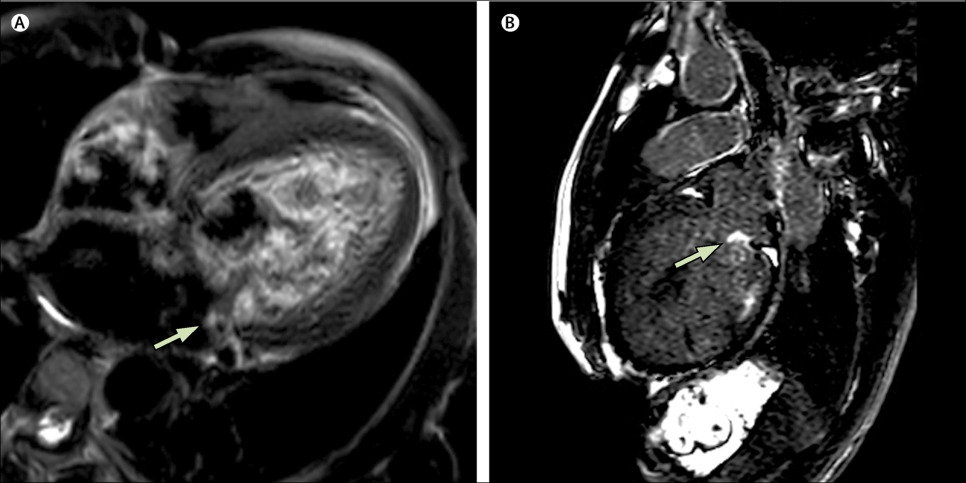
Delayed enhancement images, 4-chamber views. B2 demonstrates interventricular patchy enhancement (arrow) on cardiac MRI performed during an SLE flare and active chest pain. Appearance is typical for myocarditis and a new finding compared to the baseline scan A, with some residual involvement after resolution of the SLE flare and medical treatment (C2). The finding of myocarditis is supported by a patchy enhancement (arrow), consistent with myocardial edema and active inflammation on T2-weighted images without fat saturation. No active inflammation is noted on a followup scan (C1).
Goykman et al Subendocardial Ischemia and Myocarditis in Systemic Lupus Erythematosus Detected by Cardiac Magnetic Resonance Imaging
The Journal of Rheumatology February 2012, 39 (2) 448-450; DOI: https://doi.org/10.3899/jrheum.110812
Libman Sacks
Varied valve involvement, including valve thickening (fibrosis or calcification), leaflet perforation, stenosis, valve regurgitation and valve mass (Libman-Sacks vegetations) can be found in all valves in the heart 47. However, the mitral valve is most vulnerable.
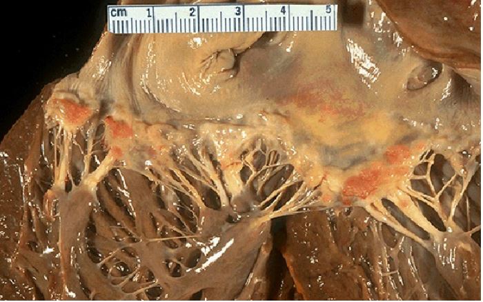
Pathology specimen shows reddish patches of thickened fibrous tissue called verrucous endocarditis formed from vegetations are small and formed from strands of fibrin, neutrophils, lymphocytes, and histiocytes. They can produce both systolic and diastolic murmurs.
The vegetations are rarely of sufficient hemodynamic importance to cause congestive heart failure.
However there is a risk of embolic disease.
Courtesy Research Gate.net
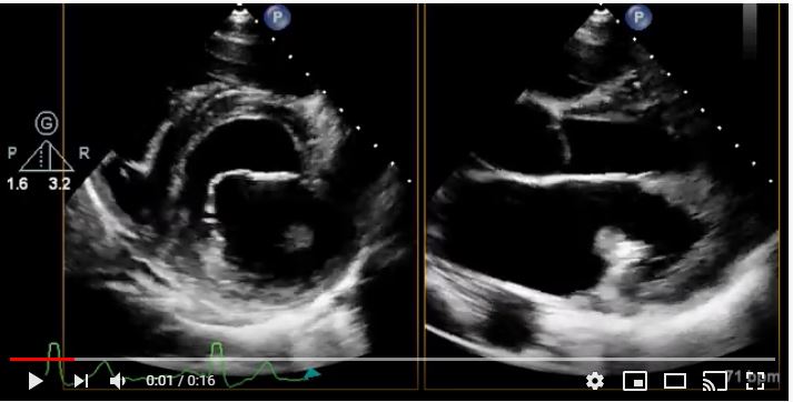
Short axis and parasternal long axis transthoracic echocardiogram, four-chamber transthoracic echocardiogram, and a zoom of a transthoracic echocardiogram all show a mass attached to the base of the posterior leaflet of the mitral valve.
Read the Clinical Picture at The Lancet: https://hubs.ly/H0ht6jJ0
Published: April 27, 2019.
Elagha et al Cardiac MRI clinches diagnosis of Libman-Sacks endocarditis
VOLUME 393, ISSUE 10182, PE39, APRIL 27, 2019
Pericarditis
Increased risk of CAD
lipid dysfunctions can accelerate the progression of overall atherosclerotic burden 4
SLE patients had a higher calcification score compared to controls
MRI
- Buzz
-
- patchy
- mid ventricular
- basal
- septal
- also vascular distribution because of vasculitis (subepicardial
-
Ntusi et al Myocardial tissue characterisation with late gadolinium enhancement in rheumatoid arthritis, systemic lupus erythematosus and systemic sclerosis
J Cardiovasc Magn Reson. 2013; 15(Suppl 1): O47.
References and Links
GoykhmanP, et al Subendocardial Ischemia and Myocarditis in Systemic Lupus Erythematosus Detected by Cardiac Magnetic Resonance Imaging The Journal of Rheumatology February 2012, 39 (2) 448-450;
Kreps, A et al Cardiac Manifestations in Systemic Lupus Erythematosus: A Case Report and Review of the Literature Am J Med Case Rep. 2018; 6(9): 180–183.
Ntusi et al Myocardial tissue characterisation with late gadolinium enhancement in rheumatoid arthritis, systemic lupus erythematosus and systemic sclerosis
J Cardiovasc Magn Reson. 2013; 15(Suppl 1): O47.