

Ashley Davidoff MD

71 year old female with long standing idiopathic pulmonary hypertension, and retroperitoneal fibrosis.
Frontal X-ray shows an enlarged triangular shaped heart and a significantly enlarged main pulmonary artery.
Ashley Davidoff MD

71 year old female with long standing idiopathic pulmonary hypertension, and retroperitoneal fibrosis.
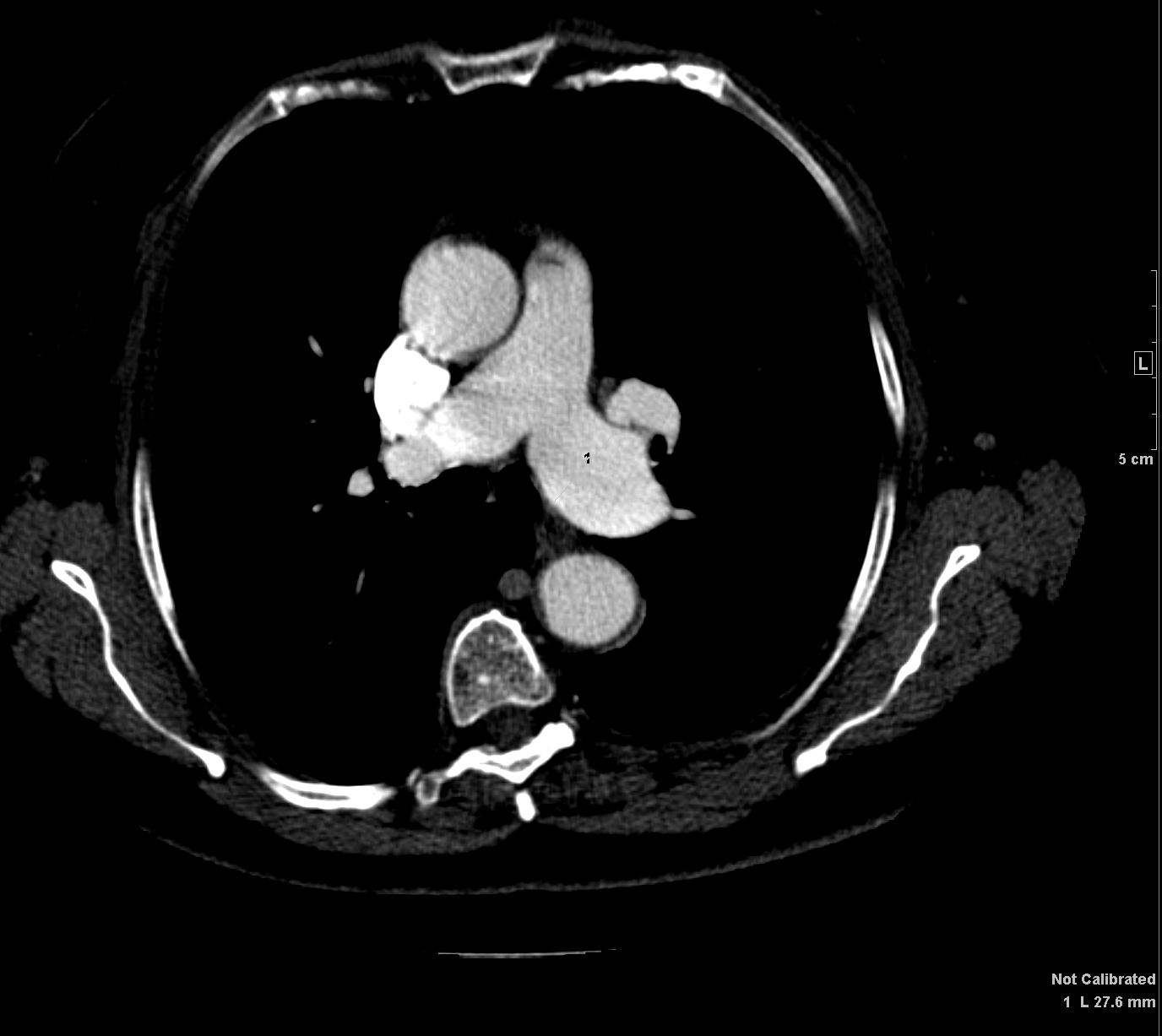
CT scan shows an enlarged main pulmonary artery (MPA) that measures 5.1cms at the level of the tubular portion of the ascending aorta.
Ashley Davidoff MD

71 year old female with long standing idiopathic pulmonary hypertension, and retroperitoneal fibrosis.
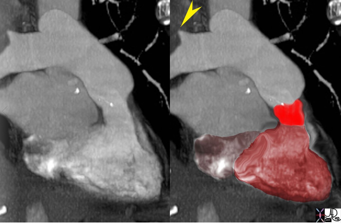
CT in the axial (a,b) coronal (c,d) and sagittal planes (e,f) show an enlarged right ventricle, (b,d,f teal arrowhead) normal sized right atrium (RA) and an atrial septum that is bulging only minimally toward the left and a significantly enlarged MPA (red arrowhead)
The MPA is enlarged. The pulmonary valve is normal in thickness, thus excluding pulmonary stenosis.
Ashley Davidoff MD
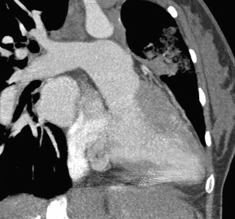
CT of patient with an anterior aorta and a large posterior pulmonary artery TGA transposition 48320 48321 DTGA
Courtesy Dr Hyun Woo Goo from Korea and Dr Laureen Sena
48321 CT of patient with an anterior aorta and a large posterior pulmonary artery TGA transposition heart aorta position 48320 48321 DTGA Courtesy Dr Hyun Woo Goo from Korea and Dr Laureen Sena
Links and References