
Ventricular Septal Defect
Copyright 2009
Section Name
Section Text
|
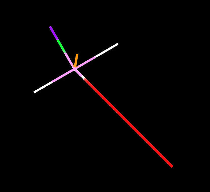
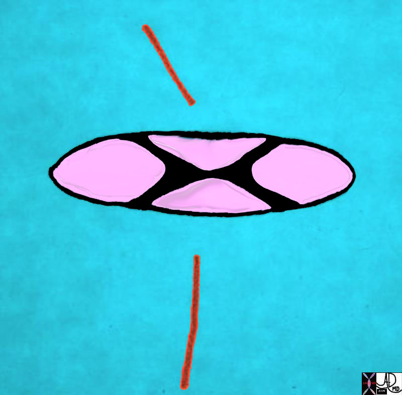
01667b14 |
| 01667b14 heart cardiac embryology conal septum line drawing right atrium left atrium left ventricle right ventricle interatrial septum interventricular septum atrioventricular septum crux of the heart embryology anatomy Davidoff drawing Davidoff MD 01667b04 01667b04 01667b06 01667b07 01667b09 01667b14 01669b03 |
|
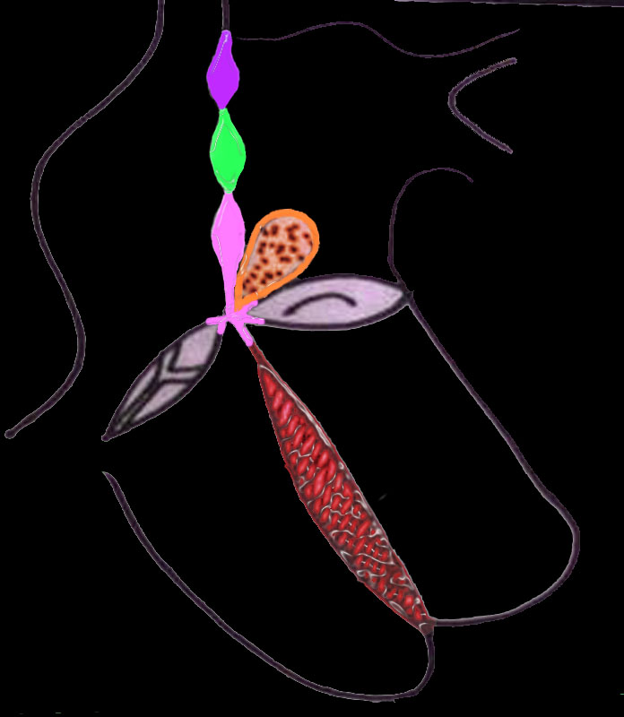
01669b03 |
| 01669b03 heart cardiac embryology conal septum line drawing right atrium left atrium left ventricle right ventricle interatrial septum interventricular septum atrioventricular septum crux of the heart embryology anatomy Davidoff drawing Davidoff MD 01667b04 01667b04 01667b06 01667b07 01667b09 01667b14 01669b03 |
|
01592c01.802 |
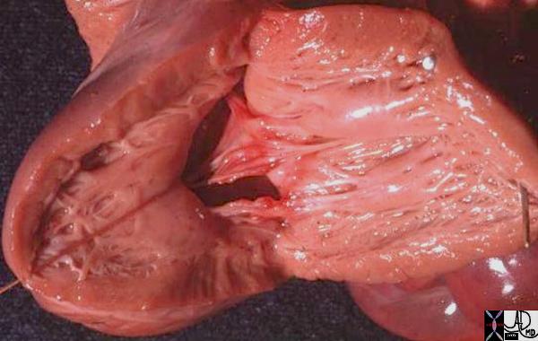
| 01592c01.802 heart cardiac mitral valve aortic valve tricuspid valve MV TV posterior leaflet septal leaflet anterior leaflet membranous septum fibrous continuity ventricular septum RVOT right ventricular outflow tract LV left ventricle RV right ventricle anatomy normal Davidoff MD |
|
06570e |
| 06570e heart cardiac atrial septum ventricular septum IVS sepatal defects ASD of primum type secundum ASD membranous VSD ventricular septal defect muscular VSD VSD of the conal septal conal VSD supracristal VSD subpulmonic VSD ASD of the AV canal type Davidoff art drawing Courtesy Ashley Davidoff MD |
|
00244b |
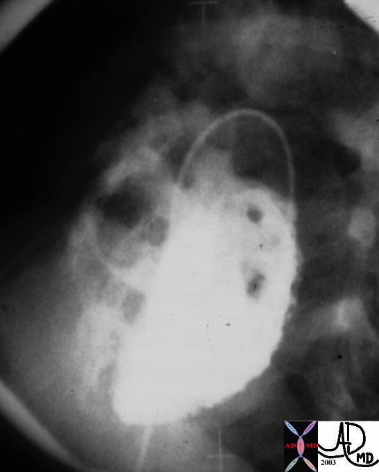
| This LV angiogram in LAO projection shows multiple ventricular septal defects with filling of the RV. The catheter course is from the aorta to the LV Courtesy Ashley Davidoff 00244b code heart LV RV interventricular septum VSD multiple muscular cardiac multiple muscular VSDs congenital imaging radiology angiography |
|
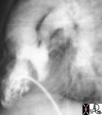
00250 |
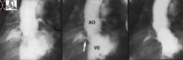
| This is a series of angiograms of the left ventricle in LAO projection showing a puff of contrast into the right ventricle through the interventricular septum. A similar collection of contrast is noted in the the main pulmonary artery just to the left of the aorta. This high VSD is in the position of the membranous septum and thus represents a mebranous ventricular septal defect.. Courtesy Ashley Davidoff MD. 00250 code cardiac heart LV RV VSD membranous MPA aorta AO congenital imaging radiology angiography |
|
00271c04 |
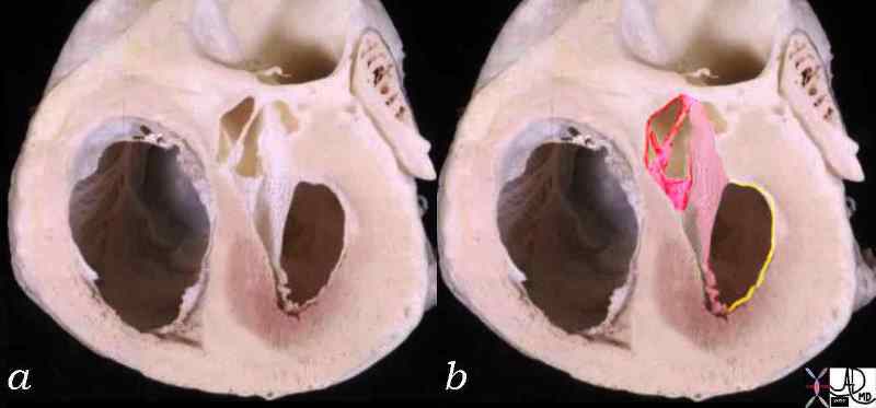
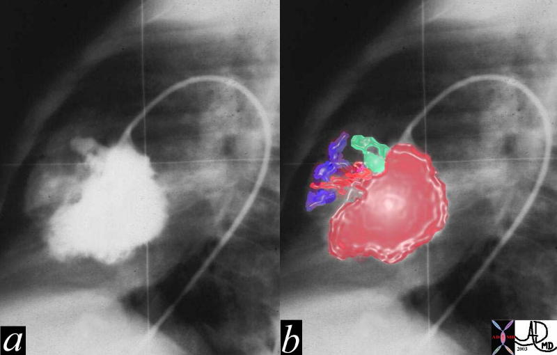
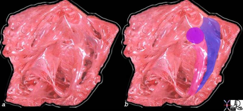
| This LV angiogram shows a high VSD at the level of the memranous septum. (a). Image b shows a green overlay of the aneurysmal changes of the VSD caused by entrapment of the VSD in the septal leaflet of the tricuspid valve. The jet of blood seems to emanate from under the valve and into the RV (blue) Courtesy Ashley Davidoff MD 00271c04 code heart cardiac VSD memranous aneurysm congenital angiography |
|
00258c02 |
| 00258c02 heart cardiac ASD secundum atrial septal defect of the primum type common atrioventricular cana’ common AVC defect superior limbic band inferior limbic band septum primum septum secundum grosspathology Courtesy Ashley Davidoff MD copyright 2008 |
|
01674b02 |
| 01674b02 heart cardiac valves endocardial cushions septum mitral valve tricuspid valve TV MV interatrial septum interventricular septum A-V canal atrioventricular canal embryology septation drawing Davidoff art Davidoff MD |
|
01863b01 |
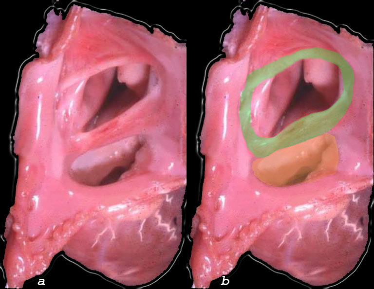
| 01863b01 heart cardiac LV left ventricle IVS interventricular septum mitral valve endocardial cushions complete AVC canal defect cleft mtral valve VSD endocardial cushion defect congenital heart disease grosspathology Courtesy Ashley Davidoff MD |
|
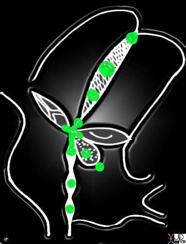
06409c02 |
| 06409c02 heart cardiac RV RVOT IVS pink = septal band purple = conal septum aka infundibular septum blue = trabeculated portion of the muscular septum tricuspid valve interventricular septum papillary muscle of Lancisi Y of septal band normal anatomy right ventricular outflow tract pulmonary valve normal anatomy grossanatomy |
|
07932c01 |
| 07932c01 heart LV cardiac defect left ventricle membranous septum ventricular septum interventricular septum VSD dacron graft repair of a membranous VSD papillary muscles grosspathology Courtesy Ashley Davidoff MD |
|
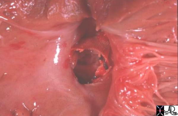
00251 |
| This pathological specimen is taken from the left ventricular chamber showing the surgical repair of a high membranous ventricular septal defect where the sutures became disrupted. Courtesy Ashley Davidoff MD 00251 code cardiac heart septum ventricular septal defect VSD post- op congenital grosspathology |
|
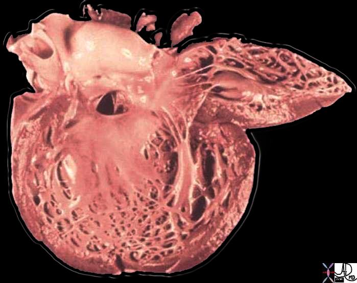
11992b03 |
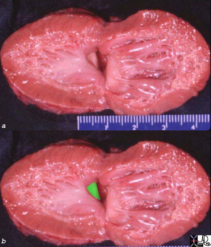
| 11992b03 heart LV left ventricle interventricular septum ventricular septum VSD ventricular septal defect subaortic grosspathology Courtesy Ashley Davidoff MD |
|
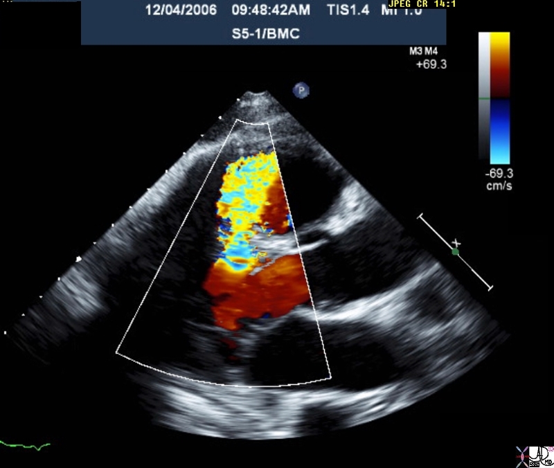
33151 |
| This color flow doppler echo of the heart with pulse flow interrogation at the ventricular septum showing high velocity turbulent flow. The patient has a diagnosis of ventricular septal defect. Courtesy Philips Medical Systems 33151 code cardiac heart echo color doppler pulse doppler high velocity LV RV VSD congenital imaging cardiac echo |
|
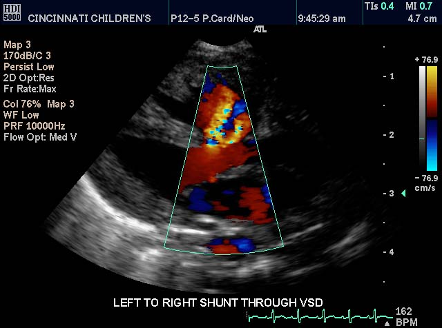
33143 |
| This color flow doppler echo of the heart shows the left ventricle anteriorly and the right ventricle posteriorly. A ventricular septal defect is demonstrated by a jet of green and red color from the LV to the RV Courtesy Philips Medical Systems 33143 code cardiac heart echo color doppler LV RV VSD congenital imaging cardiac echo |
|
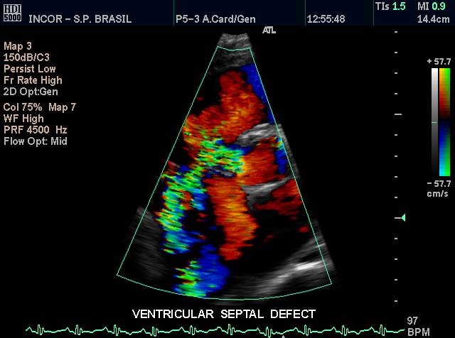
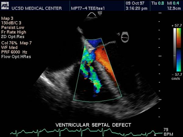
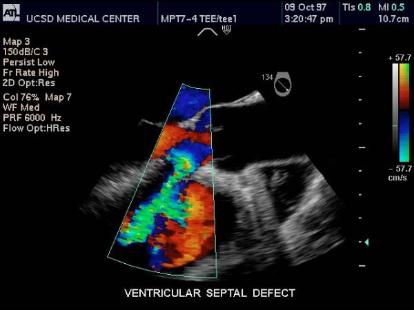
33147 |
| This color flow doppler echo of the heart showing a long axis view through the ventricles, and demonstrating a jet of turbulent color originating in the left ventricle (near view) and extending into the right ventricle. (far veiw) The patient has a diagnosis of ventricular septal defect. Courtesy Philips Medical Systems 33147 code cardiac heart echo LV RV VSD congenital imaging cardiac echo |
|
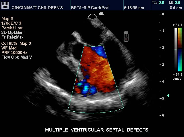
33149 |
| This transesophageal color flow doppler echo of the heart showing a 4 chamber view with the ventricles in focus show multiple jets of turbulent color extending from the apex of the left ventricle to the apical region of the right ventricular sinus. The patient has a diagnosis of multiple ventricular septal defects Courtesy Philips Medical Systems 33149 code cardiac heart echo doppler color LV RV VSD multiple muscular imaging cardiac echo congenital |
|
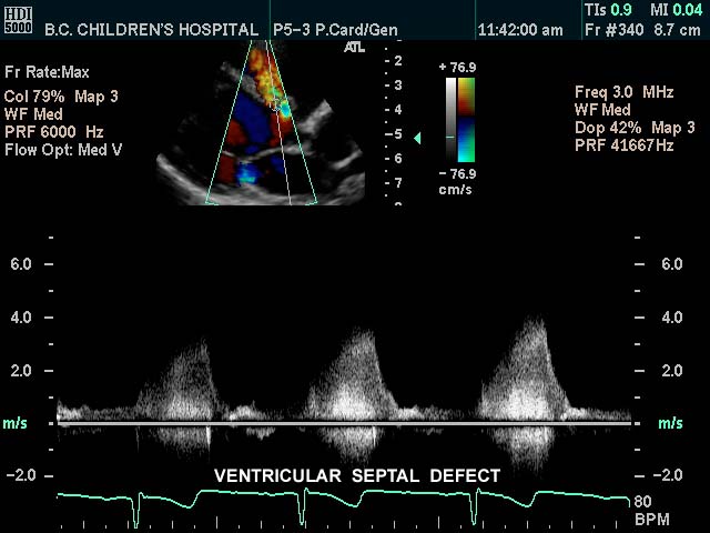
33151 |
| This color flow doppler echo of the heart with pulse flow interrogation at the ventricular septum showing high velocity turbulent flow. The patient has a diagnosis of ventricular septal defect. Courtesy Philips Medical Systems 33151 code cardiac heart echo color doppler pulse doppler high velocity LV RV VSD congenital imaging cardiac echo |
|
33174 |
| This color flow doppler echo of the heart showing a 4 chamber view, and demonstrating color flow in all the chambers at the crux of the heart at the membranous septum. This is highly suggestive of a VSD with a defect of endocardial cushions Courtesy Philips Medical Systems 33174 code cardiac heart echo LV RV RA LA VSD endocardial cushion defect congenital crux imaging cardiac echo |
|
33175 |
| This color flow doppler echo of the heart showing a long axis view, and demonstrating color jet from left ventricle to right ventricle via a ventricular septal defect that is in subaortic position. This is characteristic of a tetralogy of Fallot. Courtesy Philips Medical Systems 33175 code cardiac heart echo LV RV AO VSD tetralogy of Fallot TOFcongenital imaging cardiac echo |
|
01487 |
| This is an angiogram of the RV, showing an anteriorly placed aorta, a VSD filling the LV, and a posteriorly positioned smaller MPA. The catheter courses via the IVC into the RV. The findings are consistent with TGA and in this case a D-TGA, though it is impossible to identify the position of the aortic valve in relation to the PA in this lateral projection. An associated VSD and subpulmonary stenosis and or PS is implied by the small sized PA. Courtesy Ashley Davidoff MD 01487 code CVS heart cardiac transpoistion of the great arteries DTGA VSD subpulmonary stenosis imaging radiology angiography |
|
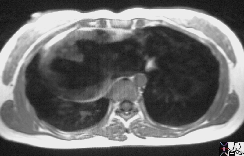
16933 |
| 16933 heart cardiac interventricular septum heart dx single ventricle dx ILR dx pulmonary atresia congenital heart disease MRI T1 weighted Courtesy Ashley Davidoff MD |
|
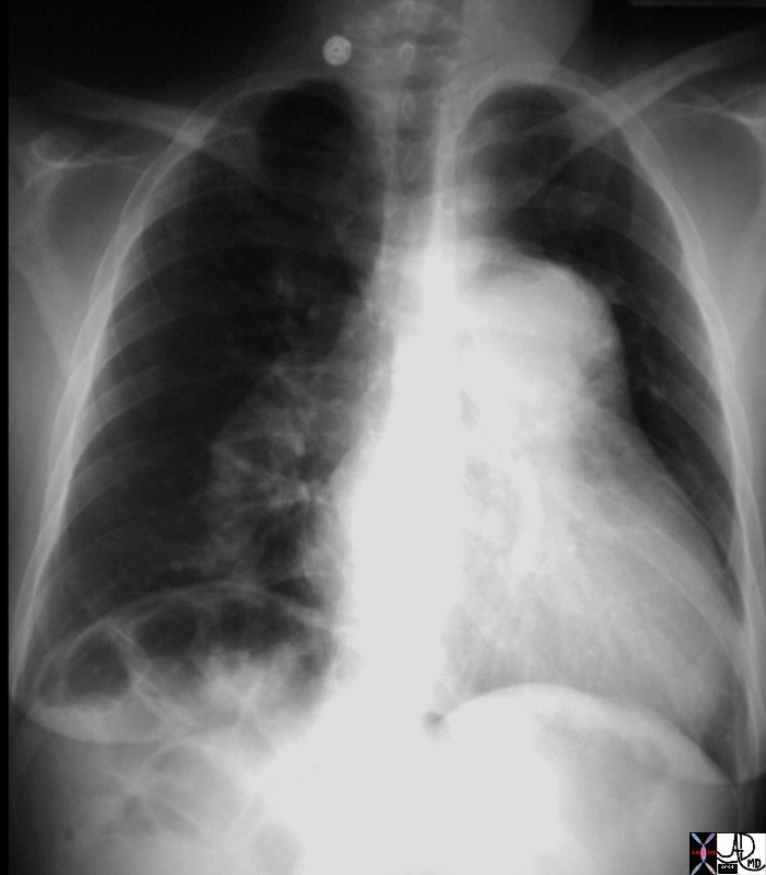
15406.800 |
| 15406.800 heart cardiac right ventricle RV fx enlarged MPA main pulmonary artery fx enlarged pulmonary arteries DD ASD VSD PDA septal defects Eisenmenger’s CXR plain X-ray of chest Davidoff MD fx pruned pruning dx Eisenmenger’s Davidoff MD 15406.800 15406c02 15406c04 15406c05 |
|
15406c04 |
| 15406c04 heart cardiac right ventricle RV fx enlarged MPA main pulmonary artery fx enlarged pulmonary arteries DD ASD VSD PDA septal defects Eisenmenger’s CXR plain X-ray of chest Davidoff MD fx pruned pruning dx Eisenmenger’s Davidoff MD 15406.800 15406c02 15406c04 15406c05 |
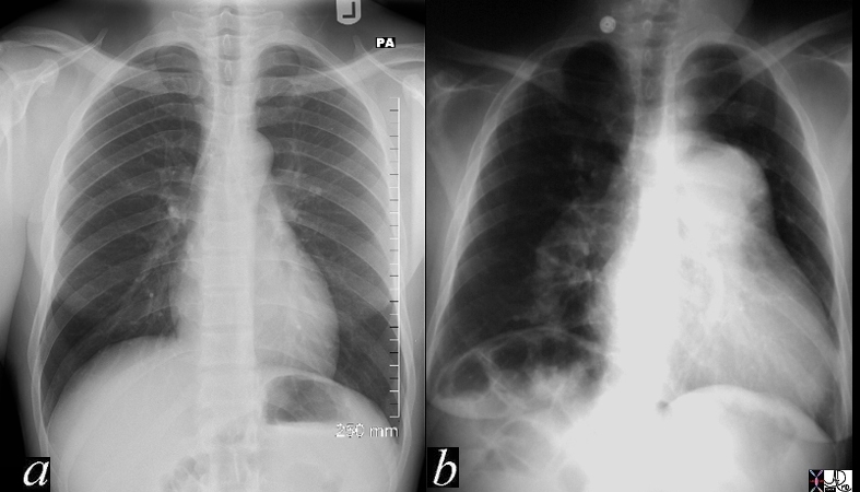
Courtesy Dr Ravin Davidoff MD
|
86780.8s |
| 86780.8s Stab wound to the heart with a traumatic VSD. The 2D echo shows a muscular VSD with turbulent left to right shunting code heart cardiac trauma septum ventricle ventricular seprum ventricular septal defect muscular septum Courtesy Ravin Davidoff MD copyright 2009 all rights reserved |
Case Studies