
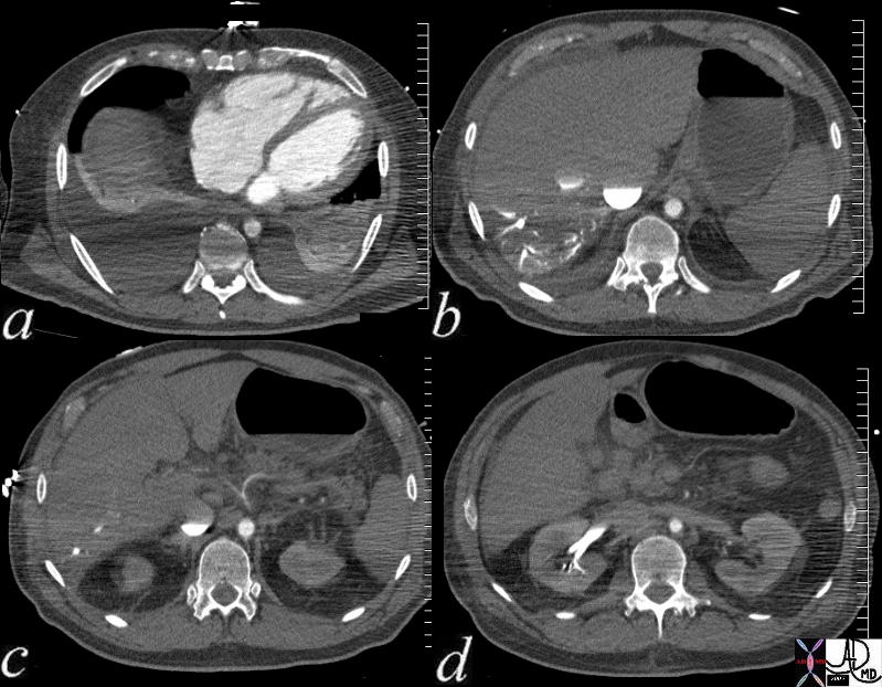
Multiple axial images through the lower chest and upper abdomen in a patient with cardiogenic shock reveals the consequences on the right side of the circulation. In image (a), the right ventricle and right atrium are enlarged and there are bilateral pleural effusions. Image (b) shows stasis of contrast into the IVC with a blood contrast level. The column is relatively static due to peripheral constriction and slow return. There is reflux into the hepatic veins due to tricuspid regurgitation and the reflux extends all the way to the periphery indicating poor forward flow in the hepatic circulation again due to peripheral constriction. Note how small the aorta is due to contraction of the muscular media in this life threatening situation. In image c the celiac axis with branches hepatic artery and splenic artery show severe vasoconstriction. In d the reflux of contrast low pressure extends deep into the renal parenchyma for the reasons outlined above.
key words
heart circulation kidney hepatic vein reflux artery celiac axis spasm aorta small shock CTscan
Courtesy Ashley Davidoff MD 73796c01