

Buzz
- Overall volume
- Shapes
- Linear Size
- Wall thickness
4 Chamber View
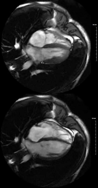
This is the MRI of a 19 year old male who presented with syncope and the study was performed to identify a possible arrhythmogenic focus
White blood imaging using 4 chamber view shows a normal sized heart in systole (above) and diastole below. The left and right atria have flattened surfaces, and occupy about 1/3 the volume of the ventricles. The RV volume is about 2/3 the volume of the LV. The wall of the LV in diastole (lower image) is less than 1 cms and the wall of the RV is barely seen and is in the range of about 3mm. Note in diastole the MV and TV are open
Ashley Davidoff MD

White blood imaging using 4 chamber view shows a normal sized heart in systole (above) and diastole (below). The left ad right atria have flattened surfaces, and occupy about 1/3 the volume of the ventricles. The A-P dimension of the LA during diastole is about 4cms (normal) and the RA is about 5cms (normal). The RV volume is about 2/3 the volume of the LV. The transverse dimension of the RV in diastole is about 4cms and the LV 5cms – both normal. The septum of the LV in diastole (lower image) is less than 9mm, and the free wall is 9mms (upper limits normal is 1.2 cms. The wall of the RV is barely seen and is in the range of about 3mm. Note in diastole the MV and TV are open. The difference in diameter of the RV in systole and diastole is about 2/3 and similarly of the LV. This an approximately normal ratio
Ashley Davidoff MD
Straight Axial View
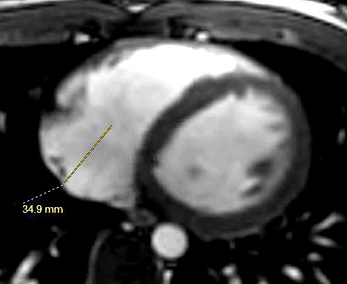
This is the MRI of a 19-year-old male who presented with syncope and the study was performed to identify a possible arrhythmogenic focus
White blood imaging of the RA, LV and RV in the axial projection is shown in diastole. The RA measures 3.5cms (n= up to about 5cms). The RA volume is about 1/3 the size of the volume of the RV.
Ashley Davidoff MD