56 year old male with history of ASD repair, alcoholism, cirrhosis post MVA with traumatic brain injury
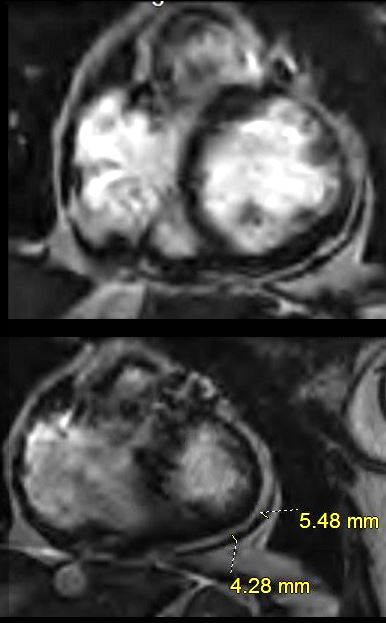
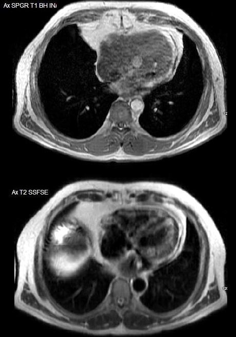
Chest X rays reveal pericardial calcification along the lateral and inferior aspect of the heart possibly due to prior surgery. MRI performed for evaluation of the cirrhotic liver to exclude hepatoma shows only a minimally irregular and thickened pericardium – up to 5mm.


56 year old male with history of ASD repair, alcoholism, post MVA with traumatic brain injury
Chest X rays reveal pericardial calcification along the lateral and inferior aspect of the heart possibly due to prior surgery.
Ashley Davidoff MD

MRI performed for evaluation of the cirrhotic liver to exclude hepatoma shows only a minimally irregular and thickened pericardium – up to 5mm
Ashley Davidoff MD

