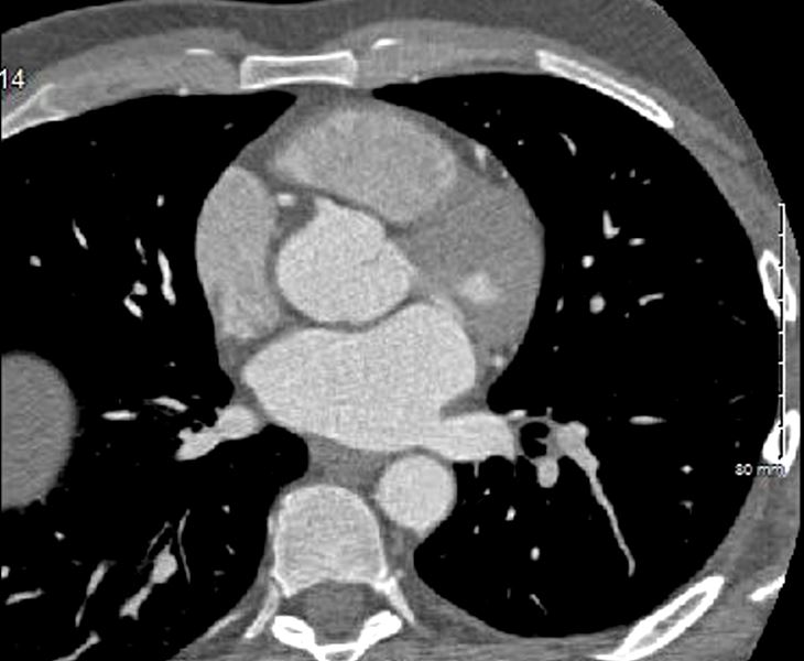
Max AP dimension of the LA in the region of the aorta. At this point the LA looks rectangular
Ashley Davidoff MD

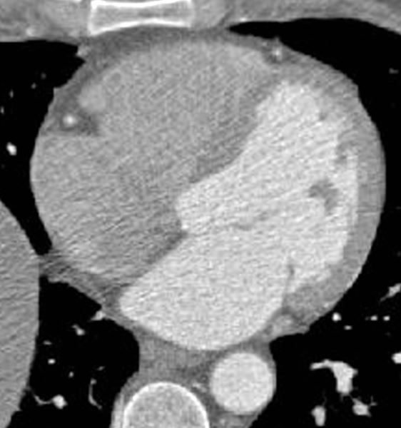
Axial images through the 4 chambers at the level of the A-V valves during diastole (mitral valve open) enables an approximate volume evaluation of the chambers. The atria are approximately the same volumes, and are about 1/3 the volume of the ventricles. The right ventricle (RV) is about 2/3 the volume of the left ventricle (LV)
Ashley Davidoff MD

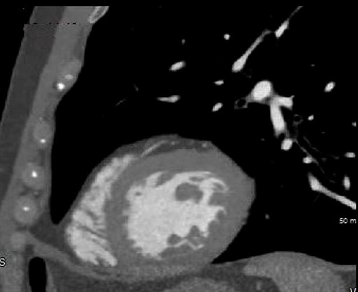
Ashley Davidoff MD 131235 sagittal 001
