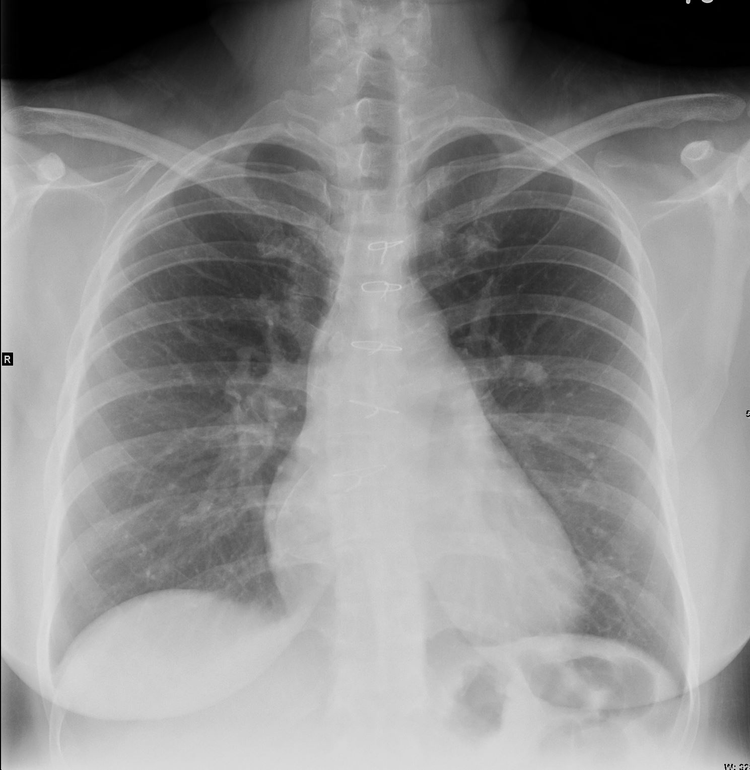
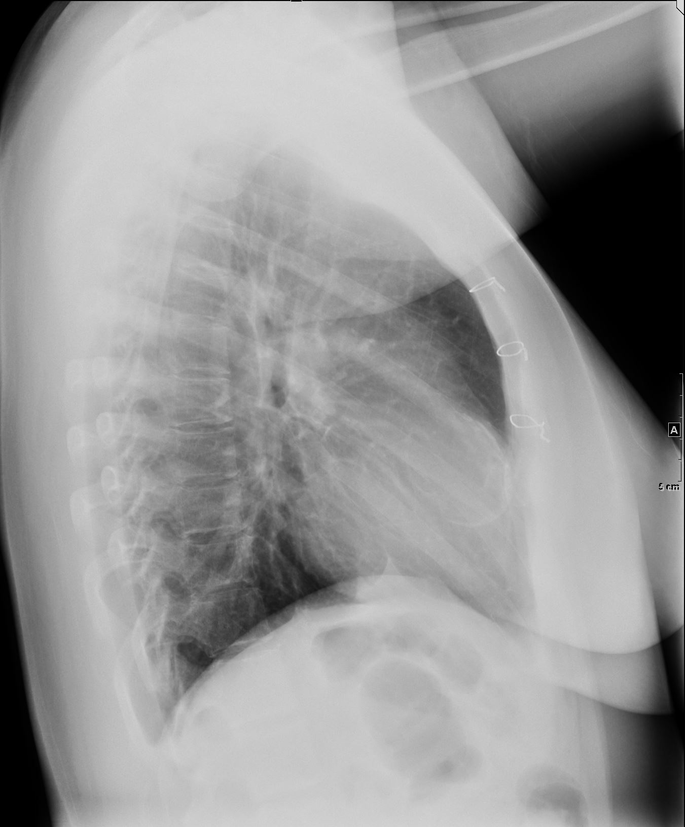
36F with RCA aneurysm iagnosed while pregnant. Seen in congenital clinic
Ashley Davidoff thecommonvein.net
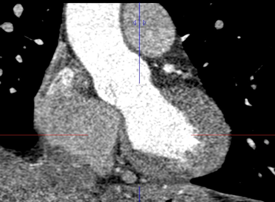
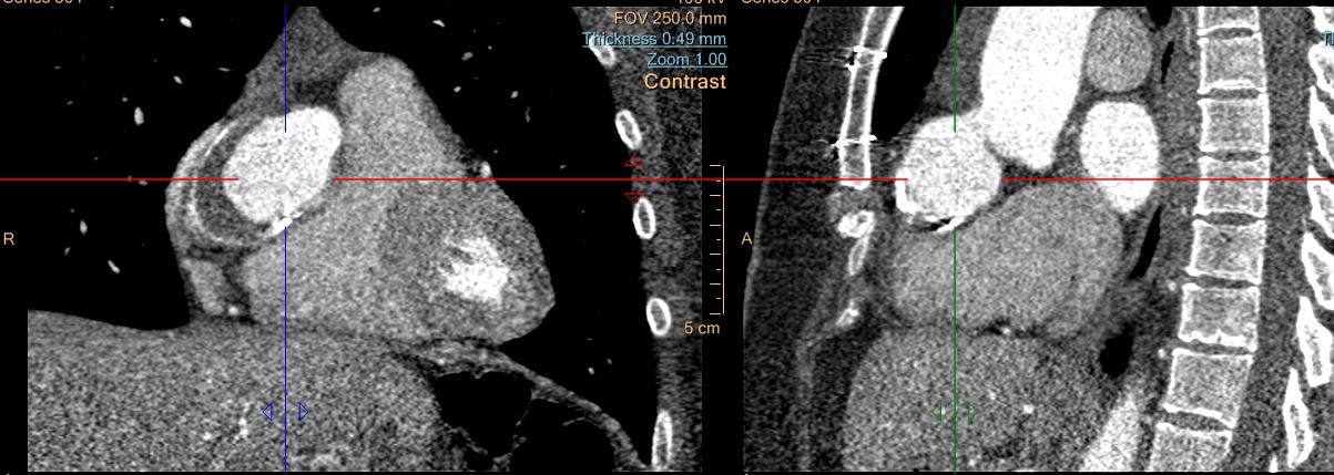
Remote RCA fistula closure (Jacksonville, FL) AngioCTCAhere with no coroanry athero; R dominant coronary circulation with normal origin. Large proximal RCA aneurysm (3.5 cm) involving ostium and spanniong 5 cnm in length. Mural thrombus within aneiurysmal sacand peripheral Ca+. Mid-distal RCA without stenosis in the RCA
CT 2016
HISTORY:
37 year old woman with history of coronary artery fisula, status post
repair as a child, now with a right coronary artery aneurysm, who
presents for follow up.
1. Large proximal right coronary artery aneurysm involving the ostium that measures up to 3.5 cm in diameter. Comparison with outside imaging is recommended to determine stability.
thecommonvein.net

thecommonvein.net

Ashley Davidoff thecommonvein.net
Ashley Davidoff thecommonvein.net
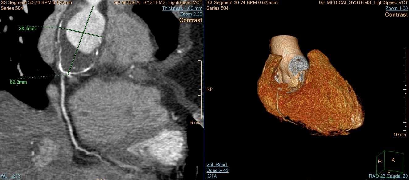
Ashley Davidoff thecommonvein.net
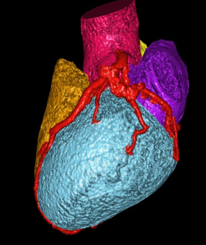
Ashley Davidoff thecommonvein.net

Ashley Davidoff thecommonvein.net
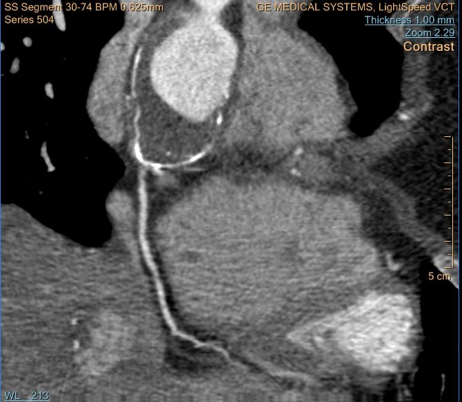
Ashley Davidoff thecommonvein.net

Ashley Davidoff thecommonvein.net

Ashley Davidoff thecommonvein.net
Latent TB
2 years ago Large proximal RCA coroanry aneurysm (3/5 cm) involving ostium and spanning 5 cm in length. Mural thrombus with aneurysmal sac and peripheral Ca++, mid-distakl RCA without stenosis I nthe RCA.
CV IMAGING: TTE (1 year ago ): LVEF 50% with Gr 1 Ddysfn Mild incr. LV wall thickness. LA/RV/RA NL. Aov and Miv OK. PV/TV NL. Ao root OK .3×3 cm round vascular lesion outside RA corresponding to the RCa aneurysm There is no IA shunt by color doppler.
UPDATE CCTA: (2 years ago ); LCA NL; LAD NL warpas around the LV apex; 2 diagonals OK ;LCX andand OMLCX OK ;RCA: ANEURYSM involving the ositium/cusp extendiong into the proximal RCAegment. 5.1×4.0x4.4 cm in current exam siht large eccentric mural thrombus and peripheral Ca++; contrast opacidified lumen 3.0×3.3 x3,3 cm and RCa ostium measuring 2.4×2,,5 cm, Mid-dstal RCa without athero or luminal narrowing. RPL and RPDA originating from RCA. Normal Aov and Miv. Normal pericardium. Normal visualized aorta. Borderline enlarged main PA @ 30 Mm./ NO CHANGES IN SIZE OF RCA ANEURYSM as comaped to study of 3 years ago
2 years ago
31-year-old female with history of RCA aneurysm since 6 years ago s/p
surgical correction of coronary AV fistula with known RCA aneurysm.
2.The The RCA aneurysm measures 5.1 x 4.0 x 4.4 cm in the current
exam and demonstrates a large eccentric mural thrombus and peripheral
calcifications, with the opacified lumen measuring 3.0 x 3.3 x 3.3
cm. There is no change in the size of the aneurysm since the prior
study of 2018
3.No significant coronary artery plaque or stenosis, except for
location of RCA aneurysm.
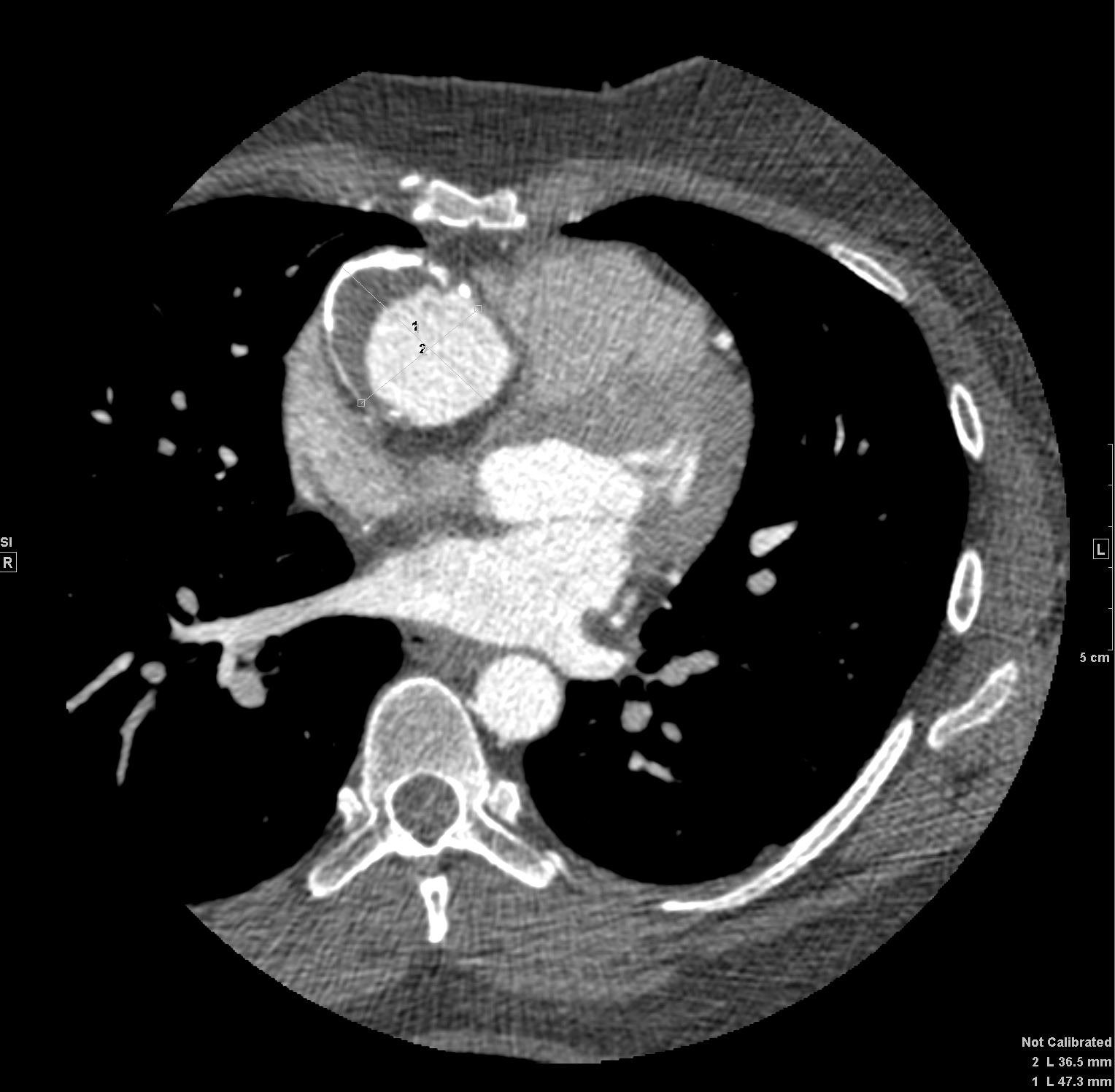
Ashley Davidoff thecommonvein.net
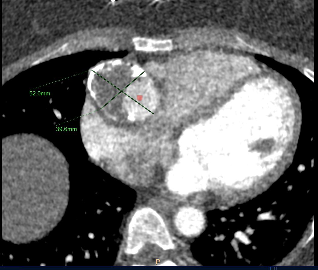
Ashley Davidoff thecommonvein.net
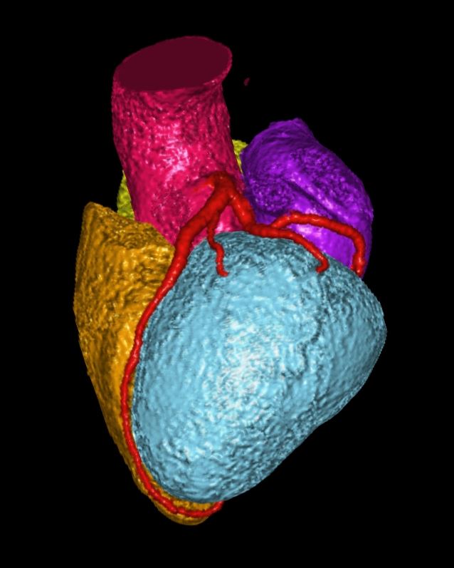
Ashley Davidoff thecommonvein.net
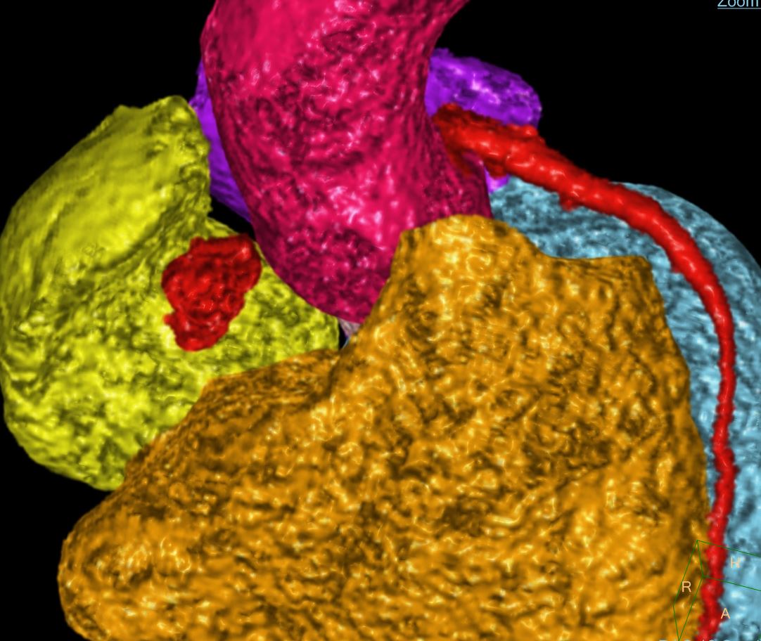
Ashley Davidoff thecommonvein.net
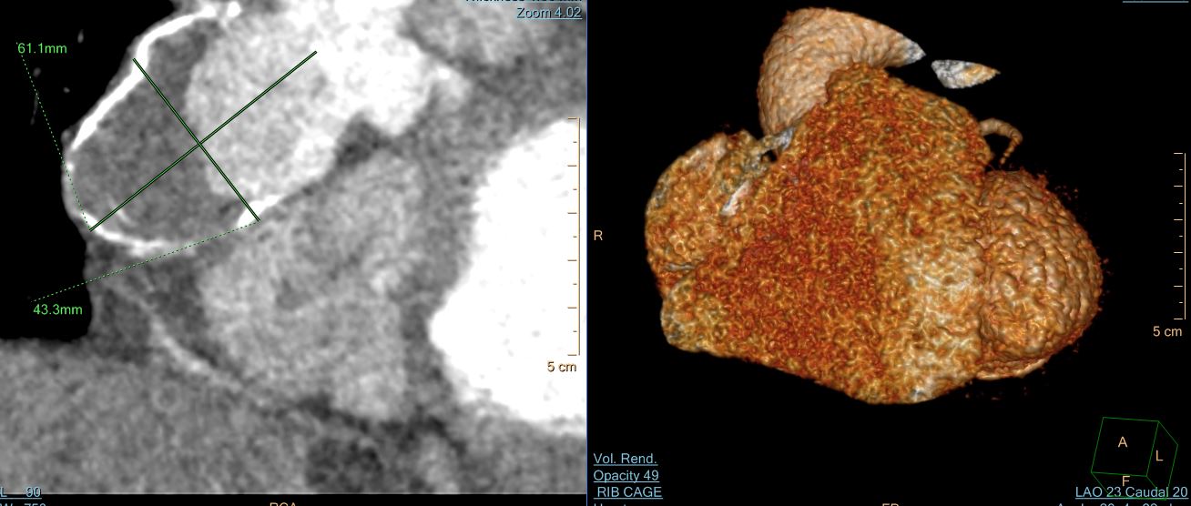
Ashley Davidoff thecommonvein.net
cURRENT
Complaint: 42 yo f. Phamacy Techniician
Ff: 1) Remote RCA fistula (Jacksonville, FL); CCTA here with no coconary atherosclerosis. Right domimnant coronary artery. LARGE PROXIMAL RCA CORONARY ANEURYSM (3.5cm , involving RCA Ostium and spanning 5 cm in length; MURAL THROMBUS within the aneurysmal sac. And, peripheral Ca++ mid to distal RCA without stneosis.
CV IMAGING: TTE (3 YEARS AGO ): LVEF 50% with Gr 1 ddysfn.normal chamber size. LA/RV/RA NL. Aortic root 3×3 cm round. Vascular lesion outsie RA correspopnding to RCA aneurysm. No interatrial shunt on color doppler.
UPDATE CCTA (2 YEARS AGO ): LCA NL. LAD wraps aroud LV Apex;and 2 diagonal branches, all OK;RCA ANEURYSM involving OSTIUM, extending into proxiomal RCA (5.1×4.0x4.4 cm with large indwelling eccentric mural thrombus and peripheral Ca++ ; contrast opacified lumen: (3.0×3.3×3.3 cm) without atherosclerosis or luminal narrowing; RPL and RPDA originating from RCA. Normal AoV and MiV and visualized ascending aorta. And pulmonary artery. NO CHANGE IN SIZE of RCA ANEURYSM compared to CCTA of 2018.
