
37 y.o. female with conoventricular VSD and bicuspid aortic valve
without significant limitations.
Past Cardiac/Medical History:
Congenital heart disease:
1- Conoventricular VSD
2- Bicuspid aortic valve (r-l fusion) with mild to moderate AR.
3- Chronic Hepatitis B s/p therapy, still with low level viremia
Family History: no CHD or SCD,
Echo
Conclusions:
Normal LV cavity size, wall thickness, and global systolic function.
Calculated LVEF is 62% using biplane modified Simpson’s method.
No regional LV wall motion abnormalities.
There is a small ventricular septal defect in the high, anterior membranous
septum (images 31, 70-75), consistent with known conoventricular VSD. There
is left to right shunt flow, but the peak velocity could not be accurately
assessed (at least 3.1 m/s, but this is likely an underestimate; systemic BP
was also not recorded).
Normal RV size and global RV systolic function.
Normal atrial sizes.
Doppler studies suggest normal LV diastolic function and normal LA pressures.
Bicuspid aortic valve with fusion of the right and left coronary leaflets,
with a raphe. Leaflets are thickened and mildly calcified. No AS. Mild to
moderate AR.
Other valves are structurally normal. There is RV outflow tract obstruction
and no PS. Trace MR. Mild TR.
Normal IVC size with blunted respirophasic variation suggestive of high
normal RA pressure.
Assuming an RA pressure of 8 mm Hg, estimated PA systolic pressure is 31 mm
Hg.
No pericardial effusion.
Compared with prior study of 10/30/2019 (images reviewed), LV size and
systolic function are similar. VSD flow is less well characterized on this
study. The degree of AR is similar.
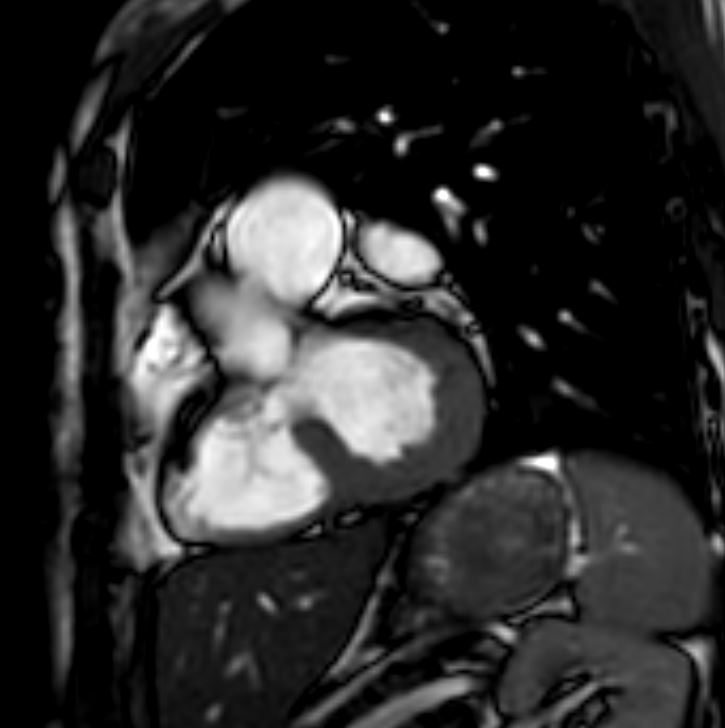
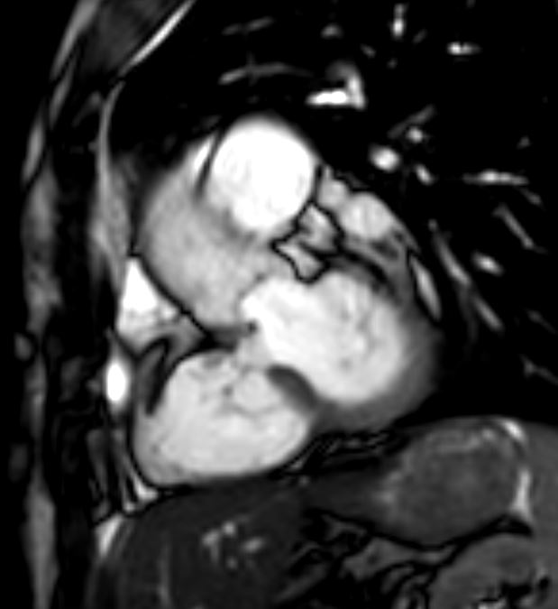
MRI
1. There is evidence of a membranous (conoventricular) VSD with a
visually small amount of left to right flow. This correlates with a QP:QS
of 1.03.
2. The LV is normal in size with normal systolic function, LVEF: 60 %.
3. The RV is normal in size with normal systolic function, RVEF: 51%.
4. No late gadolinium enhancement. Parametric analysis is normal.
5. Aortic valve leaflets are mildly thickened. Visually, there is mild
tricuspid regurgitation and mild to moderate aortic regurgitation.
6. Atria are normal sized.
7. Overall, there is a small membranous VSD with minimal left to right
flow.
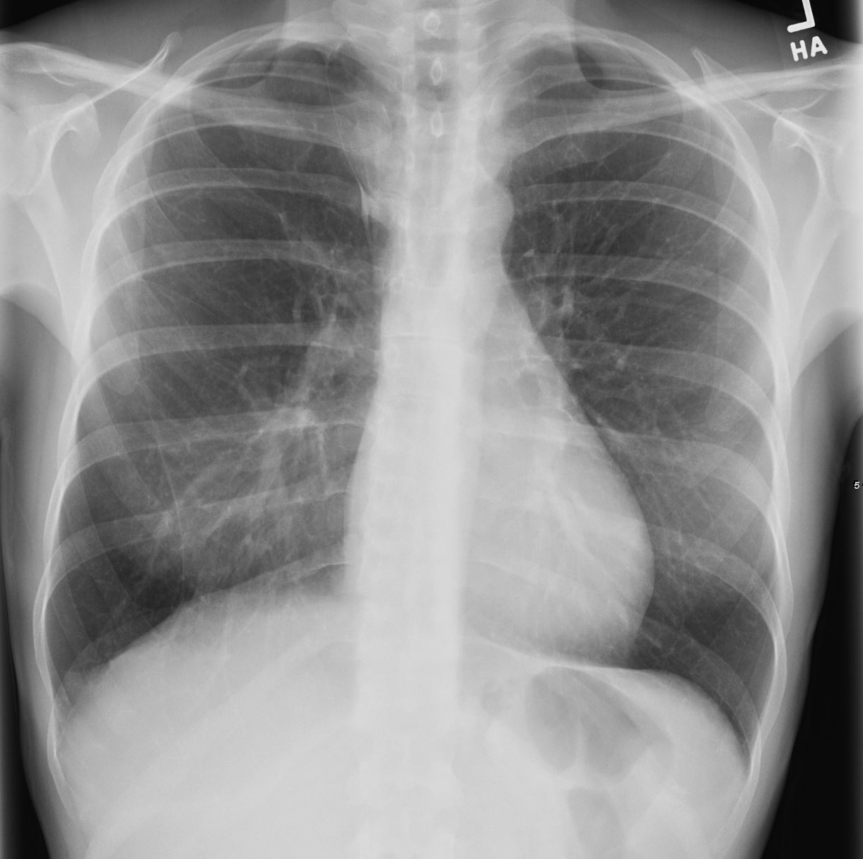
37 year old with limited symptoms
MRI shows small membranous VSD and Mild aortic stenosis with known bicuspid valve
TheCommonVein.net
Ashley Davidoff MD
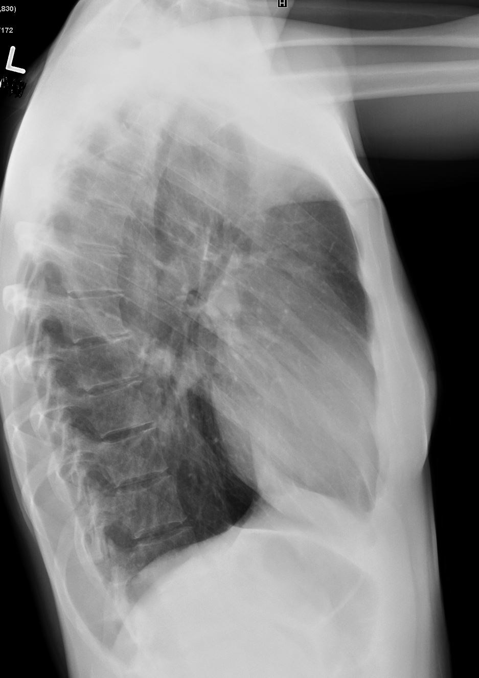
37 year old with limited symptoms
MRI shows small membranous VSD and Mild aortic stenosis with known bicuspid valve
TheCommonVein.net
Ashley Davidoff MD
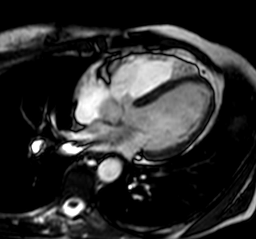
37 year old with limited symptoms
MRI shows small membranous VSD and Mild aortic stenosis with known bicuspid valve
TheCommonVein.net
Ashley Davidoff MD
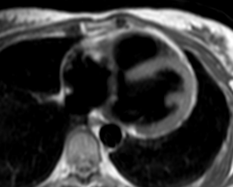
37 year old with limited symptoms
MRI shows small membranous VSD and Mild aortic stenosis with known bicuspid valve
TheCommonVein.net
Ashley Davidoff MD
37 year old with limited symptoms
MRI shows small membranous VSD and Mild aortic stenosis with known bicuspid valve
TheCommonVein.net
Ashley Davidoff MD
37 year old with limited symptoms
MRI shows small membranous VSD and Mild aortic stenosis with known bicuspid valve
TheCommonVein.net
Ashley Davidoff MD