Ventricular Septal Bulge

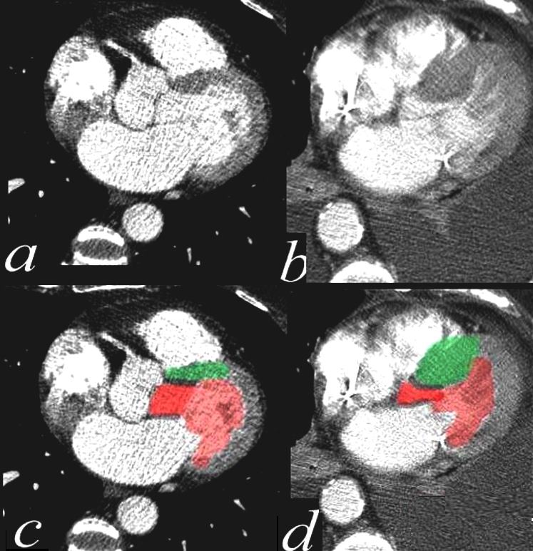
This series of CTscans shows a normal Left ventricular outflow tract overlayed in bright red in a, with normal septum in green (c) contrasted to a patient with sigmoid shaped ,septum (green in b and bright red in d) with a thickened septum with overlay in green (d). Courtesy Ashley DAvidoff MD 39208c01 code cardiac heart CTscan interventricular septum thickened IHSS imaging radiology CTscan

Ashley Davidoff
tags asymmetric subaortic septum

Ashley Davidoff
tags asymmetric subaortic septum
96-LVH-002b
VSB
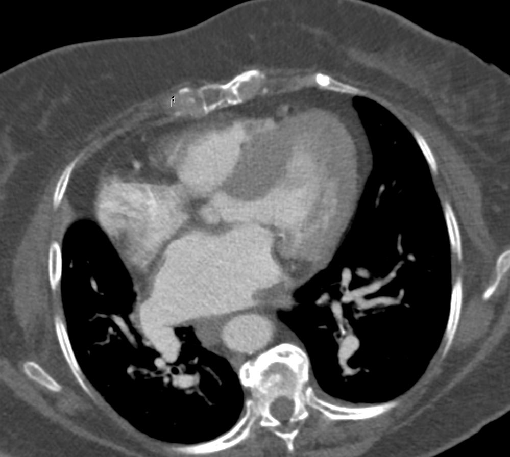
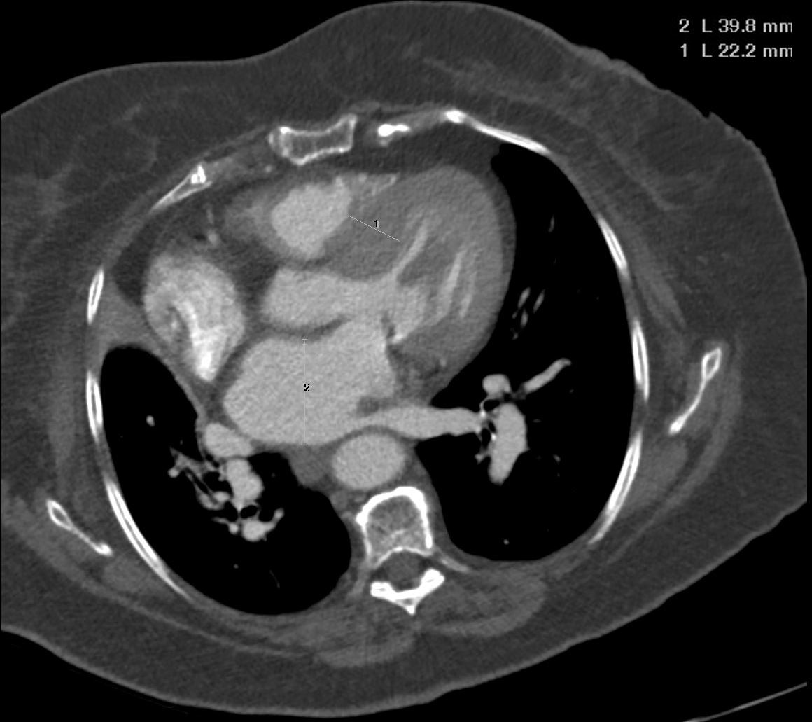
is an isolated hypertrophy of the basal segment of the interventricular septum protruding into the outflow tract of the left ventricle (LV),
- common in elderly
- 4% to 8% in individuals ≥60 years of age a
- 10% in the eighth decade of life
Hypertension
and often difficult to distinguish from genetic hypertrophic cardiomyopathy
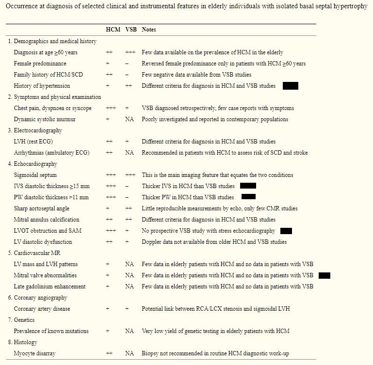
HCM is diagnosed less frequently than VSB at older ages, with a reversed female predominance. Most patients diagnosed with HCM at older ages suffer from hypertension, similar to those with VSB.
- Criteria
- isolated basal septal hypertrophy and a wall thickness >12–13 mm;
- sometimes a proximal-to-mid/distal septal wall thickness ratio ≥1.3–1.5 is used as an additional criterion,
- the presence of left ventricular outflow tract obstruction as an exclusion criterion, because considered diagnostic for HCM.
- Most patients with VSB
- Have a hx of hypertension
- between 50% and 80%
- Usually asymptomatic
- EKG
- usually no LVH
- maximal septal thickness is <15mm
- posterior wall thickness <11mm
- no mitral valve abnormalities
- Rarely have SAM and LVOT obstruction
- No LGE
- Have a hx of hypertension

Links and References
Canepa, M et al Distinguishing ventricular septal bulge versus hypertrophic cardiomyopathy in the elderly Heart. 2016 Jul 15; 102(14): 1087–1094.
