Confounding entity of indeterminate classification
May present as a morphological finding with or without clinical manifestations (CHF)
Radiopaedia “controversy as to whether non-compaction of the left ventricle represents a distinct disease versus a phenotypic manifestation of various cardiomyopathies . For example, although LVNC is classified as a primary genetic cardiomyopathy by the American Heart Association, it remains unclassified by both the World Health Organization and the European Society of Cardiology
Associated Disease
- LVOT abnormality Bicuspid aortic Valve (46%) Coarctation
- RV Disease Ebsteins (failure of RV resorption under posterior leaflet of TV) Also tetralogy of Fallot
Buzz
Non-compaction is diagnosed when the trabeculations are more than twice the thickness of the underlying ventricular wall.
- Anatomy
- infero lateral
- apex
How many n’s in the word “non”
Non Classifiable
high mortality and morbidity rates from heart failure, systemic thromboemboli, and malignant ventricular arrhythmias
Embryology

Reprinted from [46] with permission from BMJ Publishing Group Ltd.
Ikeda et al Isolated left ventricular non-compaction cardiomyopathy in adults Journal of Cardiology
Volume 65, Issue 2, February 2015
- Case 003 Non Compaction CHF and Pacemaker

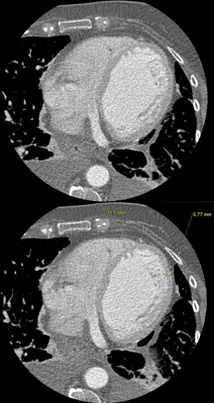
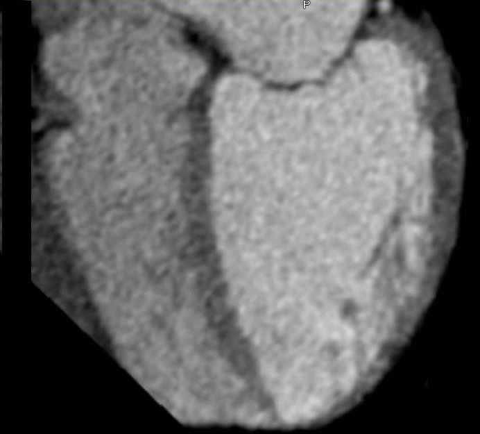
74-year-old female presents in CHF and an echo showing reduced EF (35%) and non compaction.
Initial CXR shows findings consistent with interstitial edema, (redistribution, fuzzy borders of the vessels and descending RPA) Kerley B lines, and left atrial enlargement.
Prior to implantation of a dual lead pacemaker she had a gated cardiac CT to define the venous anatomy.
The scout film shows an enlarged left atrium and suggestion of LV enlargement.
Lung windows confirmed the presence of prominent interlobular septa and LAE.
Axial soft tissue windows confirmed a diagnosis of non-compaction with non compaction thickness (NC) of 15mm and free wall thickness (C) of 6mm resulting in an abnormal NC:C ratio of 2.5 (upper limits normal NC:C ratio = 2.3)
Volume measurements showed an end diastolic volume of 217mls, an end systolic volume of 159ccs, a stroke volume of 58ccs with a resulting ejection fraction of 26%.
Ashley Davidoff MD
Case 004 Non Compaction and Ischemic Heart Disease

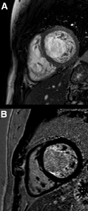
Short-axis precontrast (A) and delayed enhancement (B) views of a patient with noncompaction cardiomyopathy. On precontrast view, non-compacted layer of LV myocardium is visible. Late gadolinium enhancement sequence demonstrated LGE suggesting fibrosis of trabeculae of non-compacted layer.
Panovsky et al The prognostic impact of myocardial late gadolinium enhancement.
Cardiology in Review · May 2014

Top row: Two-chamber SSFP images in diastole (A) and systole (B) demonstrate extensive non-compacted left ventricular myocardium and apical trabeculation. Bottom row: LGE (arrows) involving the trabeculae from mid-ventricle to apex.
Mirakhur Beyond the Common Ground: The Unusual Suspects in Late Gadolinium Enhancement Conference Paper · January 2013 European Congress of Radiology
References and Links
Wan et al Varied distributions of late gadolinium enhancement found among patients meeting cardiovascular magnetic resonance criteria for isolated left ventricular non-compaction J Cardiovasc Magn Reson. 2013; 15(1): 20.
European Society of Cardiology
Wiki LV Non Compaction
TCV
