Copyright 2009
Introduction
This section is written in a specific format to conform to the principles outlined for each of the organs.
Each organ is defined by its unique structure and function, the specific diseases that affect them, the manner in which the diseases are diagnosed and the manner in which the diseases are treated.
Each structure though a unique biological unit, is part of a system that is bigger and more powerful than itself. The heart has to be connected to the other organs and systems in order to survive.
As Biological Unit
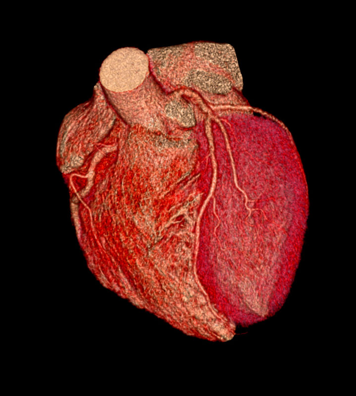
Ashley Davidoff MD
heart-0077
The heart is a hollow organ with a muscular wall that consists of inner endothelial lining, a thick muscular layer, and a double outer layer called the pericardium.
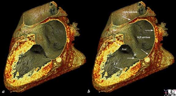
The heart is a hollow organ with a muscular wall that consists of inner endothelial lining, a thick muscular layer, and a double outer layer called the pericardium. The reconstructed CT scan shows a distended left ventricle (LV), left atrium (LA) and right atrium (RA). The thin endocardium (white arrows) throughout the heart is one continuous sheet of tissue connected across the whole circulatory system and it is more fibrous in nature. The thin white layer is seen in the left atrium, left atrial appendage (LAA) over the posterior leaflet of the mitral valve (pl) in the left ventricle (LV) and in the right atrium. The surface of the left ventricle, left ventricular outflow tract (LVOT) and right atrium are also lined by the endothelium. Ashley Davidoff, M.D.
47824c02.8s
Links and Connections
The heart has two basic connections – one to the lungs and the second to the rest of the body.
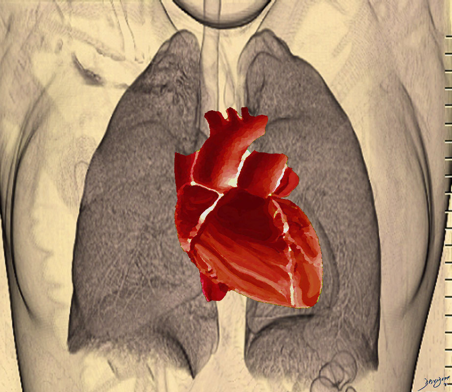
Ashley Davidoff MD
heart-0078
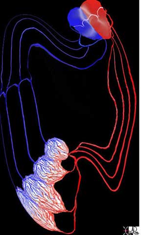
The Systemic Circulation
The heart is is intimately connected to all the tissues and cells of the body from the tip of the toes to the crown of the head.. The systemic arterial connection originates from the aorta with connections via the brachiocephalic, mesenteric, renal and peripheral arterial systems. The systemic veins join to form the inferior vena cava that drains the lower part of the body and superior vena cava that drains the upper part. The lymphatic system flows from the heart into the thoracic and right lymphatic ducts. The nerve supply is via connections to the sympathetic (celiac axis) and parasympathetic (vagus) systems. Hormonal connections include interaction with epinephrine, nor epinephrine, and thyroid hormones, but the atrial muscles also produce hormones for example atrial natriuretic peptide (ANP) that is responsible for homeostasis of water sodium potassium and adipose tissue.
Units to Unity
The cells and the chambers cannot function alone.
The cells need each other.
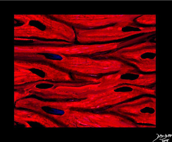
Ashley Davidoff MD
32907b03.61k.8s
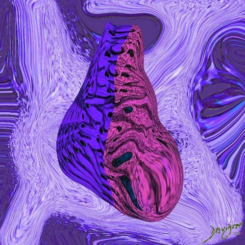
The concept of units to unity reflects the importance of each unit within the system being an important part of something larger. The heart is part of the cardiovascular system, which in turn is part of every system in the body. It is composed of the pump and its tubes – a system of vessels and capillaries in every tissue. It is a vital organ.
Dependence and Independence
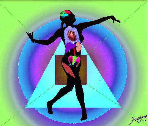
The heart cannot function alone and requires centrally based innervations and signaling of the neurohormonal axis to instruct it as to the needs of each of the other systems of the body.
Ashley Davidoff MD
The heart cannot function alone and requires centrally based innervations and signaling of the neurohormonal axis to instruct it as to the needs of each of the other systems of the body.
Time Growth and Aging
The heart arises from the primitive mesenchyme as a single tube . The single tube become a two chambered system by septation growth and resorption, and selective apoptosis. The heart ages well, though atherosclerosis of the coronary arteries and myocardial aging are normal and usual, being common causes of morbidity and mortality in the elderly.
Space
The heart lies within the thoracic cavity of the chest in close association with the lungs. It lies in an eccentric position, slightly toward the left of middle, and divides the chest into right and left sides.
Forces
The forces that govern cardiac function are mostly mechanical in origin. The forces created by contraction and relaxation allow blood to flow. The heart fills both with passive and active mechanisms by ongoing forward forces (pushing force from behind) and forces that also pull the blood (pulling forces from in front). (Visa fronte forte) Valves between the atria and ventricles and the great vessels, prevent backflow and allow forward flow into the pulmonary and systemic circulations.
Interactions
The interactions of the heart with the rest of the body are fairly simple in concept. The heart acts as the transport system of vital nutrients that are required and are vital to the function of all parts of the body. In particular it transports oxygen and carbon dioxide.The metabolic substrates are transported to the capillaries and diffuse across pressure gradients into the intercellular spaces, from where they are transported through the cell membrane. Waste products are transported in the opposite direction, from the cell into the interstitium, and then into the capillaries, and venous system to be transported to the organs of excretion.
The heart responds with beat to beat accuracy to the body needs, balances right and left circulations as well as the differential needs within the vast range of physiologic conditions from sleep to intense exercise.
States of Being – Health and Disease
By virtue of its function as a pump, diseases related to the heart are of a mechanical nature. Ischemic disease is the most common malady of the heart. When the myocardium fails, congestion occurs on the venous side and output is reduced from both pumping chambers into the arterial circulations. Often the mechanical causes of disease such as stenosis of the coronary arteries, acute coronary thrombosis, or valvular stenosis require therapies that allow for opening the blockages via mechanical, minimally invasive or surgical procedures. When the myocardial forces have been reduced to levels where function is impaired, the range of therapies relate to enhancing myocardial contractility, or reducing the work load. There is a point where conventional therapy fails and the only hope for survival is heart transplantation.
Principles of Circulation
Blood flows from high pressure to low pressure
The heart is a muscular organ and is specifically designed for transport of blood by acting as a pump. At the terminal end of the arterial circulation is a capillary system structurally designed in network formation to increase the surface area available for rapid and efficient metabolic exchange.
Tube systems are universal throughout the body. The airway, venous, lymphatic, ductal, gastrointestinal, and genitourinary systems are classical examples. Flow will only occur when a pressure difference exists between the two sides of the tubes.
The velocity of flow relates to diameter, resistance, friction, and pressure. Velocity of flow will also depend on whether the flow is laminar or turbulent. If it is laminar, it will be governed by Poiseuille’s law which states that laminar flow is directly proportional to the driving pressure. Thus if the driving pressure is doubled, the flow rate will be doubled, as long as the radius remains constant. The radius of the tube, however, is very important and relates to the velocity to the 4th power. Hence if the radius of a tube is increased by 2 cm, the velocity will increase by a factor of 16, as long as all the factors remain constant including the presence of laminar flow. As velocity increases, however, the flow tends to become turbulent. Flow tends to be laminar when the tubes are small and turbulent when the tubes are large or when there are irregularities in the walls.
Resistance of flow depends on sympathetic tone of the smooth muscle. In health this tone is well balanced with the needs of the person. However, if the tone is increased due to autonomic effects to the extent that it limits adequate blood flow, ischemia manifests with pain.
The cardiovascular system is beautifully designed to enable rapid and efficient delivery of blood to the organs. Not only is the delivery rapid, but the design is such that differential needs are accommodated. Thus during exercise, the skeletal muscle is hyperactive while the gastrointestinal tract is not active. Thus blood is shunted to the skeletal muscle and away from the gastrointestinal tract.
What a system! If we could only have such a system in the world that delivers adequate amount of supplies efficiently to the regions that most need the substrates at a particular moment in time.
< prev next >
