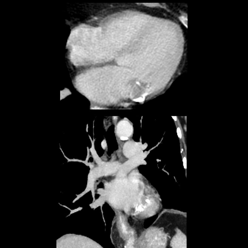
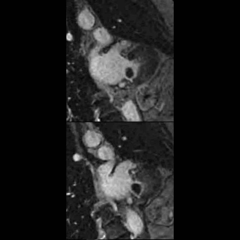
69year old female with MAC who presents with an echo finding of a mass on the mitral valve thought to represent a myxoma. CT confirmed a low-density mass measuring in the soft tissue range associated with mitral annular calcification. MRI showed a mass in the region of the posterior leaflet of the MV with T2 hyperintensity suggesting aqueous component and T1 hyperintensity suggesting increased protein content. There was no enhancement of the lesion. Peripheral T1 and T2 low intensity reflects calcification. A diagnosis of caseous necrosis of MAC is most likely
Ashley Davidoff MD
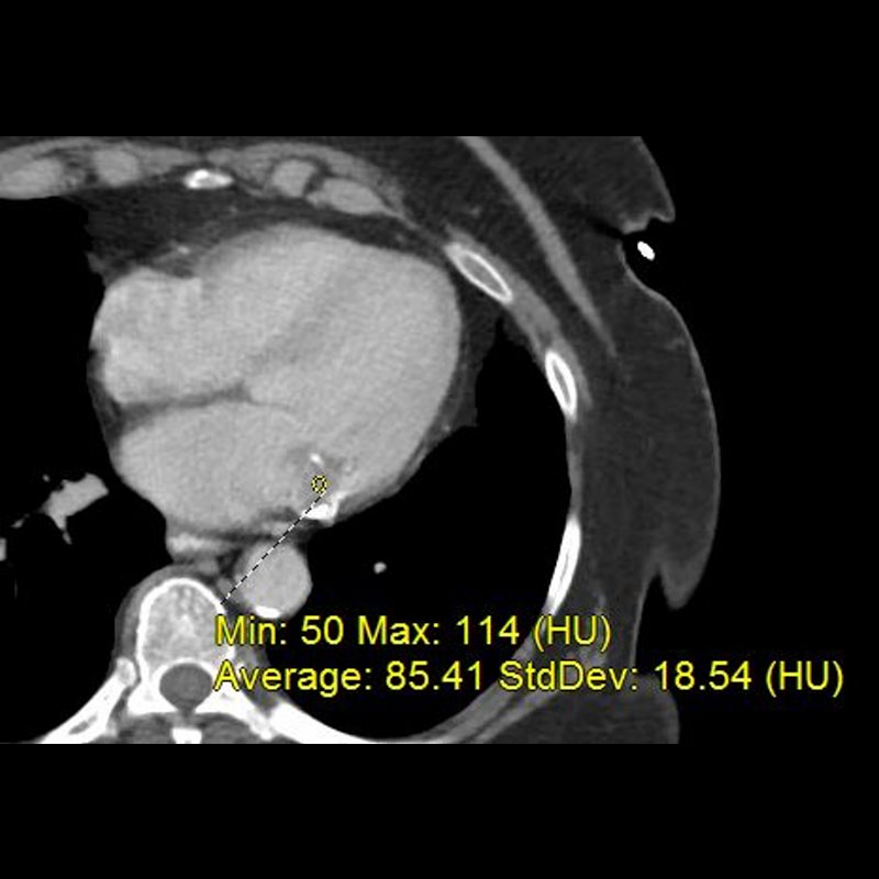
69 year old female with MAC who presents with an echo finding of a mass on the mitral valve thought to represent a myxoma. CT confirmed a low-density mass measuring in the soft tissue range associated with mitral annular calcification. MRI showed a mass in the region of the posterior leaflet of the MV with T2 hyperintensity suggesting aqueous component and T1 hyperintensity suggesting increased protein content. There was no enhancement of the lesion. Peripheral T1 and T2 low intensity reflects calcification. A diagnosis of caseous necrosis of MAC is most likely
Ashley Davidoff MD
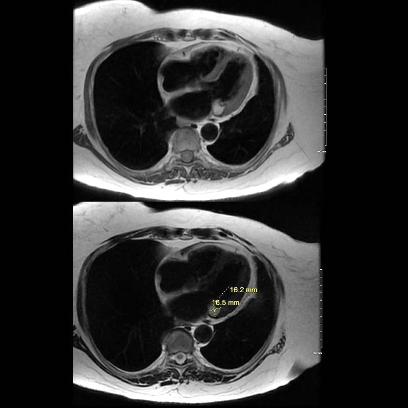
69 year old female with MAC who presents with an echo finding of a mass on the mitral valve thought to represent a myxoma. CT confirmed a low-density mass measuring in the soft tissue range associated with mitral annular calcification. MRI showed a mass in the region of the posterior leaflet of the MV with T2 hyperintensity suggesting aqueous component and T1 hyperintensity suggesting increased protein content. There was no enhancement of the lesion. Peripheral T1 and T2 low intensity reflects calcification. A diagnosis of caseous necrosis of MAC is most likely
Ashley Davidoff MD
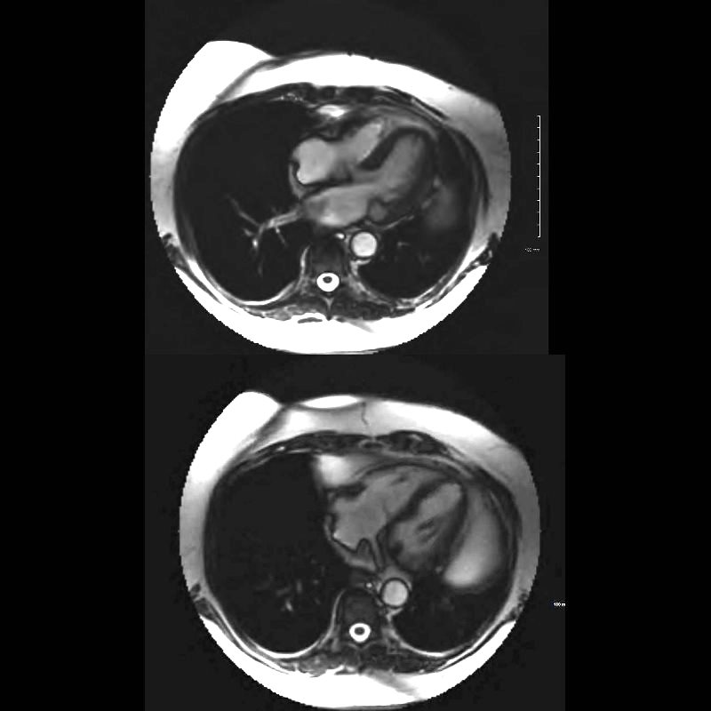
69 year old female with MAC who presents with an echo finding of a mass on the mitral valve thought to represent a myxoma. CT confirmed a low-density mass measuring in the soft tissue range associated with mitral annular calcification. MRI showed a mass in the region of the posterior leaflet of the MV with T2 hyperintensity suggesting aqueous component (upper image) and focal T1 low intensity nodule on the posterior leaflet reflects the MAC. There was no enhancement of the lesion. A diagnosis of caseous necrosis of MAC is most likely
Ashley Davidoff MD
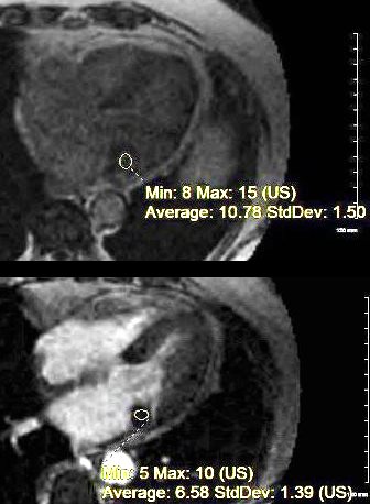
69 year old female with MAC who presents with an echo finding of a mass on the mitral valve thought to represent a myxoma. CT confirmed a low-density mass measuring in the soft tissue range associated with mitral annular calcification. MRI showed a mass in the region of the posterior leaflet of the MV with T2 hyperintensity suggesting aqueous component and T1 hyperintensity suggesting increased protein content. There was no enhancement of the lesion. Peripheral T1 and T2 low intensity reflects calcification. A diagnosis of caseous necrosis of MAC is most likely
Ashley Davidoff MD

