The Common Vein
Ashley Davidoff MD 2020
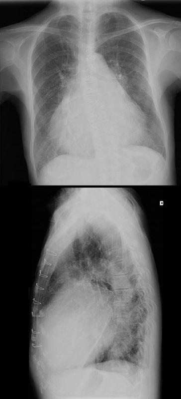
71 year old Asian female with rheumatic heart disease dominated by calcific mitral stenosis mild MR, moderate tricuspid regurgitation and secondary pulmonary hypertension.
Ashley Davidoff MD
In the P-A Projection
INCREASE CARDIOTHORACIC RATIO – MITRAL STENOSIS PULMONARY HYPERTENSION AND COR BOVINUM
71 year old Asian female with rheumatic heart disease dominated by calcific mitral stenosis mild MR, moderate tricuspid regurgitation and secondary pulmonary hypertension.
Ashley Davidoff MD
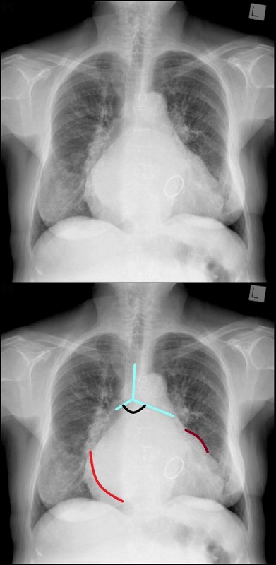
The frontal CXR demonstrates findings consistent with mitral stenosis including a widened carinal angle (teal blue and black arc), a double density (red arc) and an enlarged left atrial appendage (maroon arc).
The overall shape of the heart is triangular suggesting right ventricular enlargement. A mitral valve prosthesis is in position
Courtesy of Radiopaedia
|
01818.6s |
| 01818.6s The anatomical specimen is from a patient with mild hypoplastic left heart syndrome requiring aortic valve replacement. There was only mild mitral valve disease but there was significant aortic stenosis requiring valve replacement. The chordae in this case are thickened and foreshortened though the papillary muscle are well developed. heart LV left ventricle mitral valve MS AS aortic valve stenosis HLHS hypoplastic left heart syndrome gross pathology Courtesy Ashley Davidoff copyright 2008 all rights reserved |
35 year old patient with severe mitral stenosis . with left atrial enlargement (LAE) characterized by elevation of the left mainstem bronchus, straightening of the left heart border, and prominence of the upper 1/3 of the posterior border of the heart. The right atrium is also enlarged characterized by a rotund right heart border. There is right ventricular enlargement characterized by filling in of the retrosternal airspace.
Ashley Davidoff MD
|
19729 |
| This chest CT through the middle of the left ventricle (LV) shows some calcification athe base of the mitral valve (MV) focal calcification of the anterior leaflet and and increase density of both leaflets. This patient had no known mitral valve disease and the findings most likely represent dystrophic calcification due to mucinous degeneration. Courtesy Ashley Davidoff MD. 19729 code heart mitral valve MV calcification cardiac imaging radiology CTscan |
|
19730 |
| 19730 heart pleural calcification cardiac interventricular septum IVS LV mitral valve calcification rheumatic heart disease probable chronic hemothorax or empyema anemia fx relatively dense interventricular septum due to anemia CTscan Courtesy Ashley Davidoff MD |
|
35315 |
| 35315 Courtesy of Laura Feldman MD. code calcification enlarged heart LA LAE mitral MS MV stenosis valve cardiac heart disease imaging radiology CXR plain film |
|
32113 |
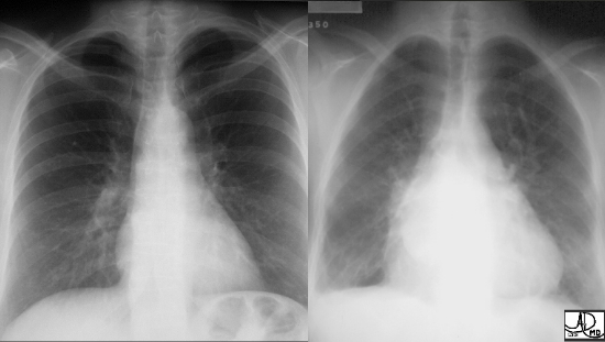
| Two patients showing the normal cardiac silhouette on the left image and an enlarged triangular shaped heart with left atrial enlargement in a patient with mitral stenosis. Notice how the apex of the heart in the second image is turned upward. Courtesy of Ashley Davidoff M.D. 32113 code cardiac heart normal disease LAE RVE imaging radiology CXR plain film |
|
32114 |
| The “double density” that we discussed above is a classical sign of an LA enlargement. The enlarged LA is in red overlay with its right sided border shifted rightward accounting for the double density. The combination of an enlarged RV and enlarged LA is most commonly seen in mitral valve stenosis. Courtesy of Ashley Davidoff M.D. 32114 |
|
34299 |
| 34299 heart cardiac MV mitral valve fx thickened LAE enlarged left atrium thickened chordae paillary muscle complex fx enlarged DDX IHD rheumatic cardicitis RHD Libman Sacks rheumatic heart disease CTscan Davidoff MD 34305 34306 34298 34299 34300 |
|
Mitral and Aortic Stenosis |
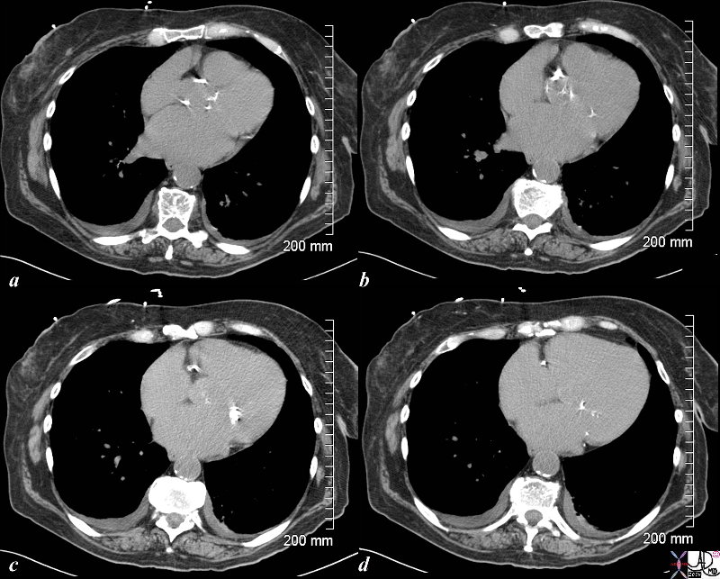
| The CT scan shows both aortic valve calcification (a,b) and mitral valve calcification (c,d) indicative of both aortic stenosis and mitral stenosis. This is likely caused by rheumatic heart disease.
76655c02.8s heart cardiac mitral valve MV MS mitral stenosis calcification calcified valve aorta aortic valve aortic calcifications CTscan Courtesy Ashley Davidoff MD copyright 2009 all rights reserved |
|
Cor Bovinum |
|
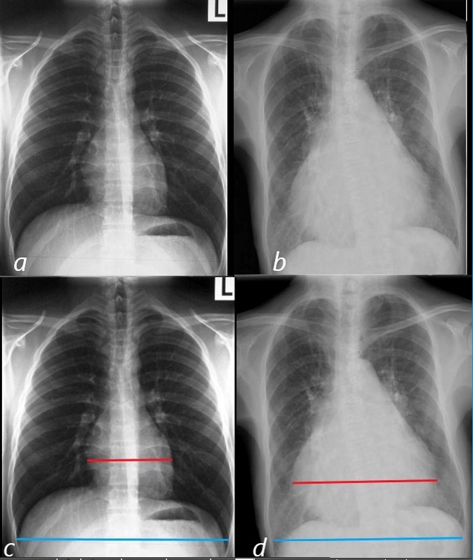
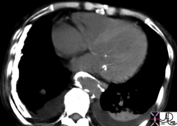
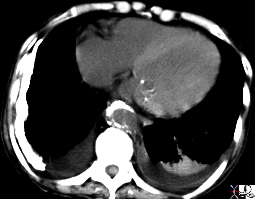
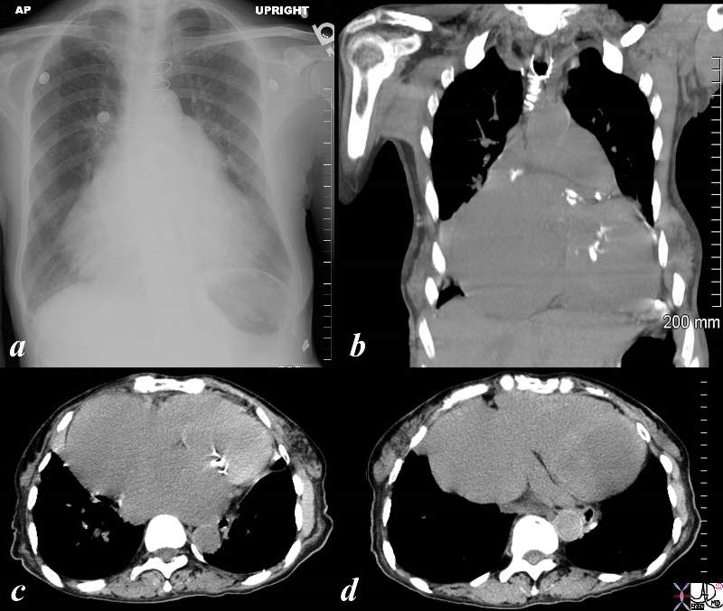
The series of a plain film of the chest (a), and coronal image of reconstructed CT (b), reveals an almost wall to wall heart called a cor bovinum – literally a bulls heart – corresponding to its very large size. The axial images c, show calcified mitral valve consisted with mitral stenosis, secondary left atrial enlargement and pulmonary hyperttension and extremely dilated right atrium seen in both image c, and d. Courtesy Ashley Davidoff copyright 2009 all rights reserved 86859c02.8s |
|
33170 |
| This gray scale echo of the heart showing a 4 chambered view with left ventricle and mitral valve in focus. The mitral valve is thickened. The patient has a diagnosis of bacterial endocarditis. Courtesy Philips Medical Systems 33170 code cardiac heart echo MV thick bacterial endocarditisinfection SBE imaging cardiac echo |
|
76286c05 |
||
76286c05 elderly lady with dyspnea heart cardiac left atrium LA calcified LA s/p Carpentier rings in mitral valve tricuspid valve and AVR aortic valve replacement rheumatic heart disease RHD mitral stenosis fx interstitial lung disease ILD reticulonodular pattern dx probable pulmonary hemosiderosis with calcified or ossified nodules scout CXR CTscan Courtesy Ashley Davidoff MD
|
|
06704c01.8s |
| 06704c01.8s This anatomical specimen is from an unfortunate pediatric patient who had a mitral valve replaced and an ASD secundum closed. The pink overlay in b shows a suture line opposing the borders of the ASD secundum. A prosthetic mitral valve is noted. heart mitral valve atrial septal defect secundum repair treatment surgery Courtesy Ashley DAvidoff MD copyright 2008 all rights reserved |
|
72794c01 |
| 72794c01 lung chest MVR mitral valve replacement hemopneumothorax pacemaker cardiomegaly CXR X-ray chest Courtesy Ashley Davidoff MD |
|
76286c04 |
| 76286c04 elderly lady with dyspnea heart cardiac left atrium LA calcified LA s/p Carpentier rings in mitral valve tricuspid valve and AVR aortic valve replacement rheumatic heart disease RHD mitral stenosis dx probable pulmonary hemosiderosis with calcified or ossified nodules scout CXR CTscan Courtesy Ashley Davidoff MD |
Links and References
Cases TCV

