Copyright 2008
Definition
Rheumatic heart disease is an autoimmune sequela to beta-hemolytic streptococcal infection of the pharynx. When the heart is involved the most frequently affected valve is the mitral valve and then most frequent is the aortic valve.
Rheumatic mitral stenosis, mitral regurgitation, aortic stenosis or aortic regurgitation results
Structurally there is thickening of the mitral valve or aortic cusps, commissural fusion and thickening and shortening of the chordae tendine. Functionally there is a narrowing of the valve orifice that impedes the free flow In MS there is progressive rise of left atrial pressure (LAP) to promote normal transmitral flow volume, while in AS there is progressive afterload on the LV. MR causes volume overload on the LA and if severe compromises LV output. AR increases end diastolic volume, and when severe decease stroke volume. Combination of aortic and mitral valve disease can be devastating.
Clinically patients present with exertional dysnea , orthopnea and PND. Auscultation reveals a characteristic. For MS there is a mid diastolic low-pitched, rumbling murmur best heard with the bell of the stethoscope with an openening snap and presystolic accentauation. In AS there is a diamond shaped ejection systolic murmur. In MR there is a pansystolic murmur, while in AR there is an early diastlic rumble .
Diagnostically, 2D Echocardiography is the diagnostic study of choice . Doppler echocardiography quantifies the hemodynamic severity of mitral stenosis .
Treatment modalities for mitral stenosis include either minimally invasive percutaneous ballon valvotomy or surgical commissurotomy, open or closed . Aortic valve replacement is usually required for severe aortic disease.

Mitral Stenosis |
| Two patients showing the normal cardiac silhouette on the left image and an enlarged triangular shaped heart with left atrial enlargement in a patient with mitral stenosis. Notice how the apex of the heart in the second image is turned upward.
Courtesy of Ashley Davidoff M.D. 32113 code cardiac heart normal disease LAE RVE imaging radiology CXR plain film |

Mitral Stenosis – Left Atrial Enlargement |
| The “double density” that we discussed above is a classical sign of an LA enlargement. The enlarged LA is in red overlay with its right sided border shifted rightward accounting for the double density. The combination of an enlarged RV and enlarged LA is most commonly seen in mitral valve stenosis.
Courtesy of Ashley Davidoff M.D. 32114 |

Rheumatic Heart Disease – Aortic Valve Mitral Valve and Tricuspid Valve |
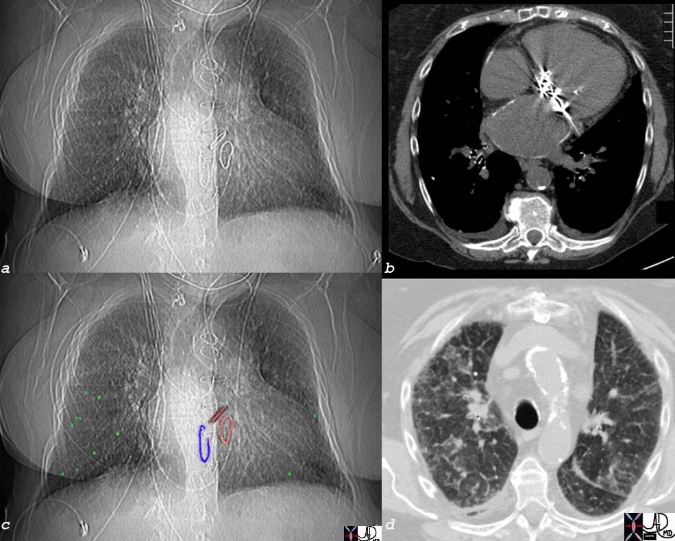
| This elderly lady had rheumatic fever as a child and developed both mitral and aortic disease that required valve replacement. As a result of the left sided diseases, pulmonary hypertension and tricuspid regurgitation resulted and she required a Carpentier ring to maintain competence of the tricuspid valve. Image a shows the three rings of metallic density in mitral aortic and tricuspid position. Image b shows a prosthesis in the mitral annulus and a calcified wall of the left atrium. Image c shows calcified nodules (overlaid in green) likely representing pulmonary hemopsiderosis, while image d shows a coarse reticular pattern and a calcified nodule in the anterior segment of the left upper lobe..
76286c05 elderly lady with dyspnea heart cardiac left atrium LA calcified LA s/p Carpentier rings in mitral valve tricuspid valve and AVR aortic valve replacement rheumatic heart disease RHD mitral stenosis fx interstitial lung disease ILD reticulonodular pattern dx probable pulmonary hemosiderosis with calcified or ossified nodules scout CXR CTscan Courtesy Ashley Davidoff MD |
 
Rheumatic Mitral Stenosis |
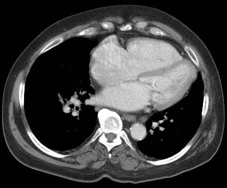
| Calcification and thickening of the anterior leaflet of the mitral valve is characteristic of rheumatic mitral valve disease.
34299 heart cardiac MV mitral valve fx thickened LAE enlarged left atrium thickened chordae paillary muscle complex fx enlarged DDX IHD rheumatic cardicitis RHD Libman Sacks rheumatic heart disease CTscan Davidoff MD 34305 34306 34298 34299 34300 |

Rheumatic Heart Disease |
| 76286c04 elderly lady with dyspnea heart cardiac left atrium LA calcified LA s/p Carpentier rings in mitral valve tricuspid valve and AVR aortic valve replacement rheumatic heart disease RHD mitral stenosis dx probable pulmonary hemosiderosis with calcified or ossified nodules scout CXR CTscan Courtesy Ashley Davidoff MD |
 
Rheumatic Heart Disease – Focal Apical Aneurysm |
| 33510 34306 heart cardiac LV apex left ventricle fx calcification calcified coronary sinus fx enlarged DDX IHD rheumatic cardicitis embolism to LAD CTscan Davidoff MD 34305 34306 34298 34299 34300 |
