Copyright 2009
“Atherosclerotic aneurysms are usually multiple and involve more than one coronary artery as compared with congenital, traumatic, or dissecting aneurysms that are often solitary. The RCA is the most frequently involved vessel (40-61%), followed by the LAD (15–32%), and circumflex coronary artery” (15–23%). El Quindy
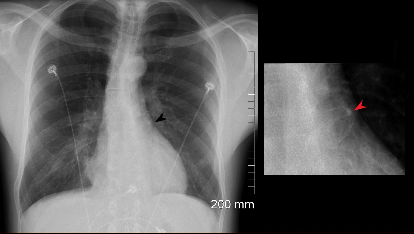
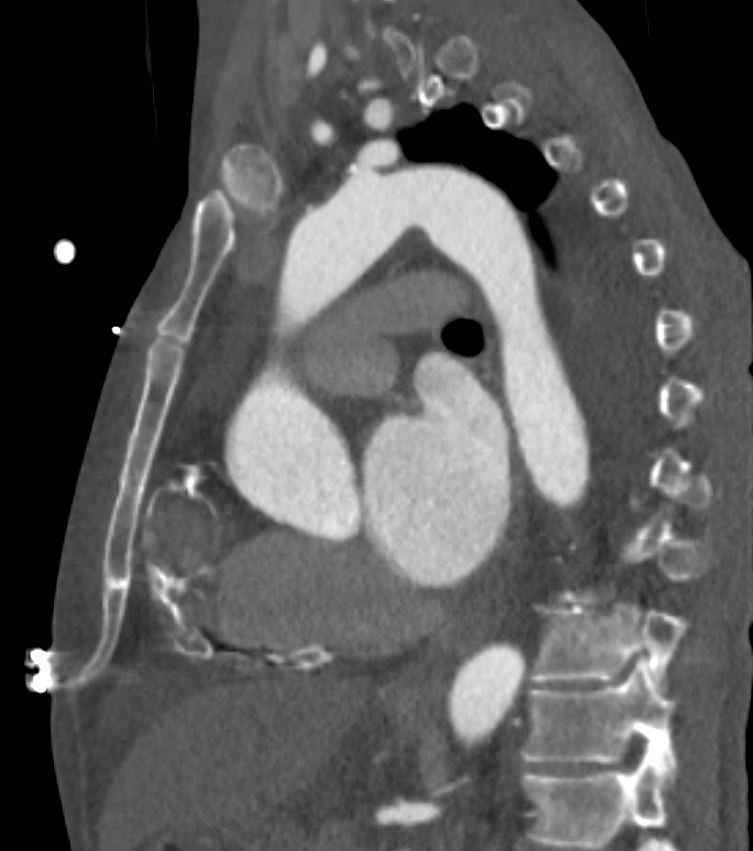
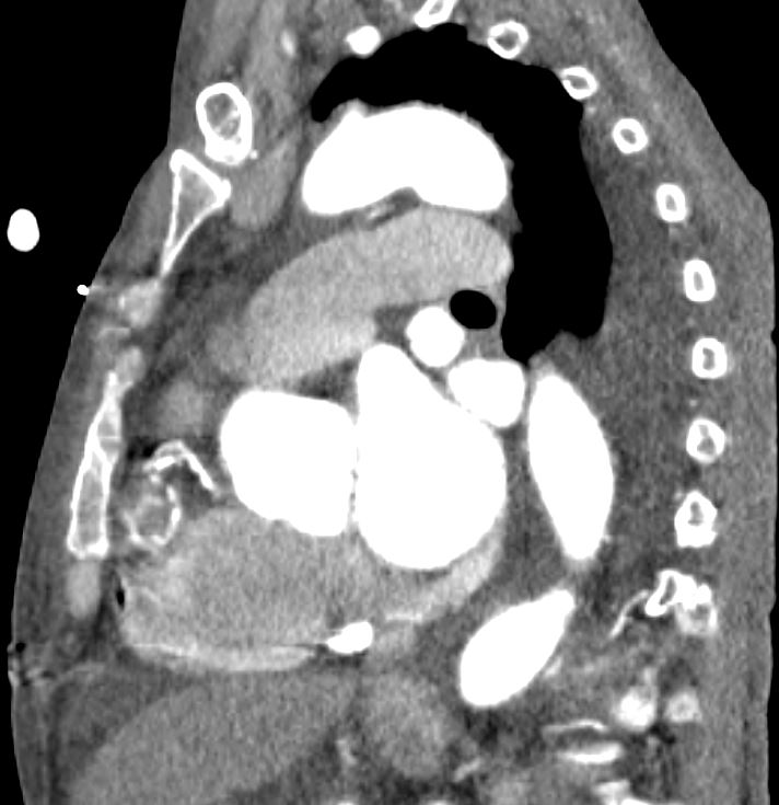
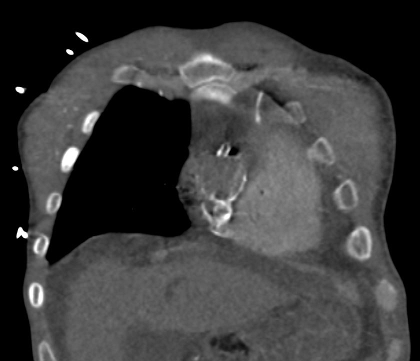
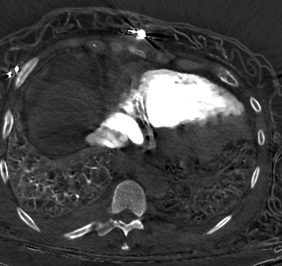
41 year old male presents with chest pain.
Frontal CXR shows a rounded calcification, (black arrow in the left image and red arrow in the magnified right image). The calcification is approximately 2cms in size and represents an aneurysm of the left coronary artery (LCA).
Ashley Davidoff MD
86928.8c
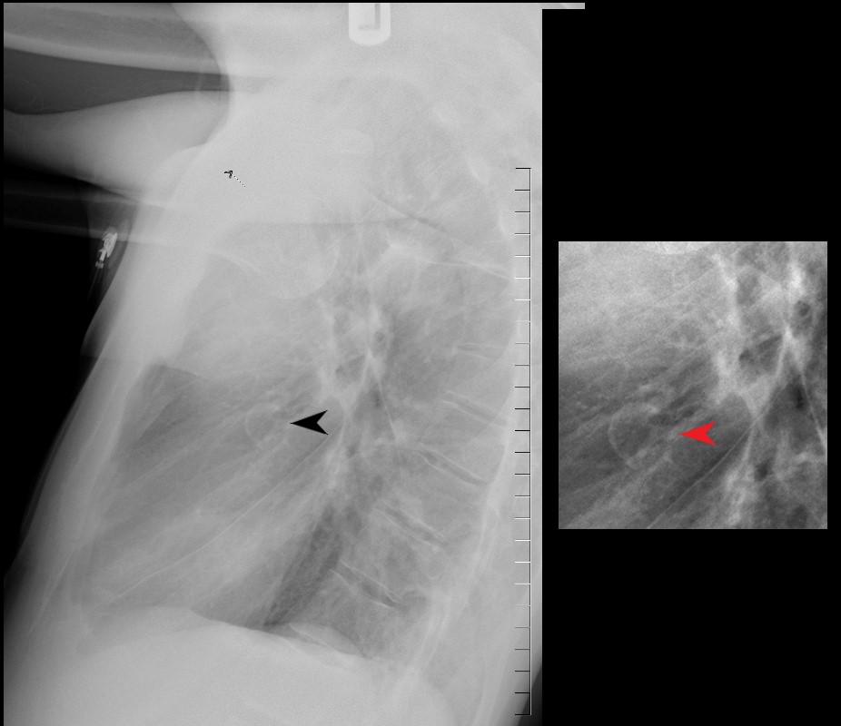
41 year old male presents with chest pain.
Lateral CXR shows a rounded calcification, (black arrow in the left image and red arrow in the magnified right image). the calcification is approximately 2cms in size .
Ashley Davidoff MD
86928.8c01
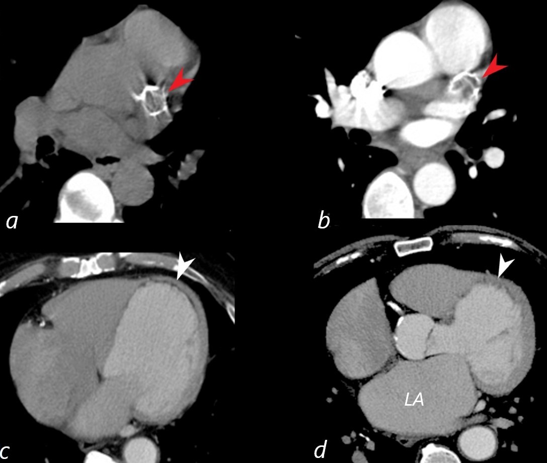
41 year old male presents with chest pain.
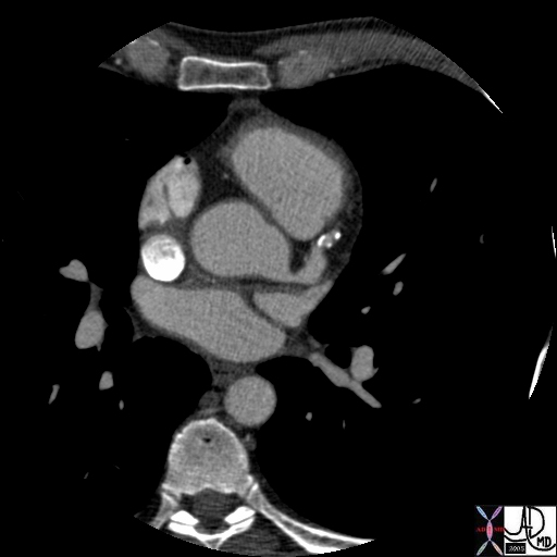
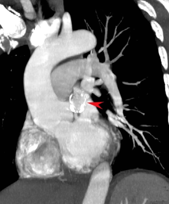
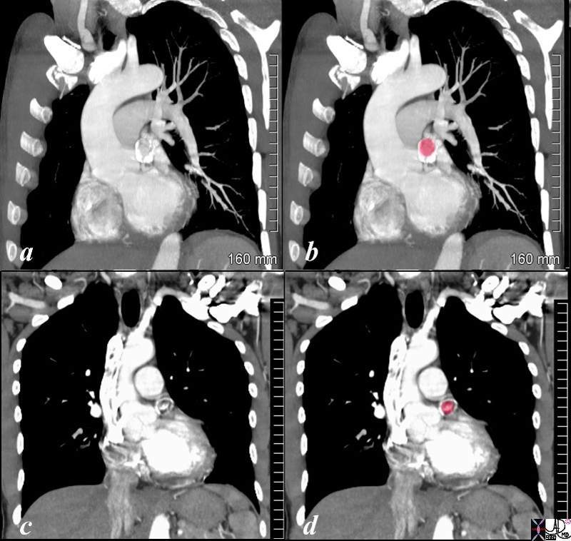
Axial CT without contrast (a) reveals aneurysmal calcification in the region of the proximal LAD,(red arrow). In image b is a similar image following contrast administration. The aneurysm is mostly thrombosed (red arrow). The calcification is approximately 2cms in size and represents a thrombosed aneurysm of the left coronary artery (LCA).
Imagesof the left ventricle ( c,d ) show a dilated LV, with subendocardial fatty infiltration of the LV myocardium in the septum, and apex (white arrowhead). There is thinning of the myocardium in the septum and apex.
These findings are consistent with myocardial infarction with and ischemic cardiomyopathy.
The left atrium (LA) is enlarged as noted in image d.
Ashley Davidoff MD
86943c01L01
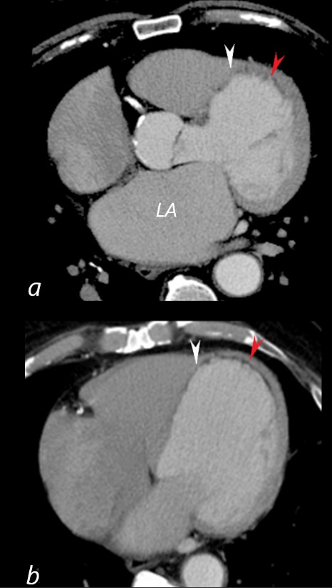
41 year old male presents with chest pain.
Axial CT with contrast at the level of the enlarged left atrium (LA) (a) reveals a dilated LV, with subendocardial fatty infiltration of the LV myocardium in the septum (white arrowhead), and apex red arrowhead). There is thinning of the myocardium in the septum and apex.
Image B is more inferior and taken at the level of the dilated RA and normal sized RV showing similar subendocardial fatty infiltration of the LV myocardium in the septum (white arrowhead), and apex red arrowhead). There is thinning of the myocardium in the septum and apex. The transverse dimension of the LV was 7.2 cms which is significantly dilated
These findings are consistent with myocardial infarction with and ischemic cardiomyopathy.
Ashley Davidoff MD
86943c03L
Ashley Davidoff MD
86923L
.
Courtesy Ashley Davidoff
86945c01.8s
|
Aneurysmal Coronary Artery Disease |
| 07614 heart cardiac aneurysms aneurysmal atherosclerosis stenosis occluded LAD with reconstitution via collaterals coronary artery angiogram angiography Courtesy Ashley Davidoff MD |
|
Aneurysmal Coronary Artery Disease |
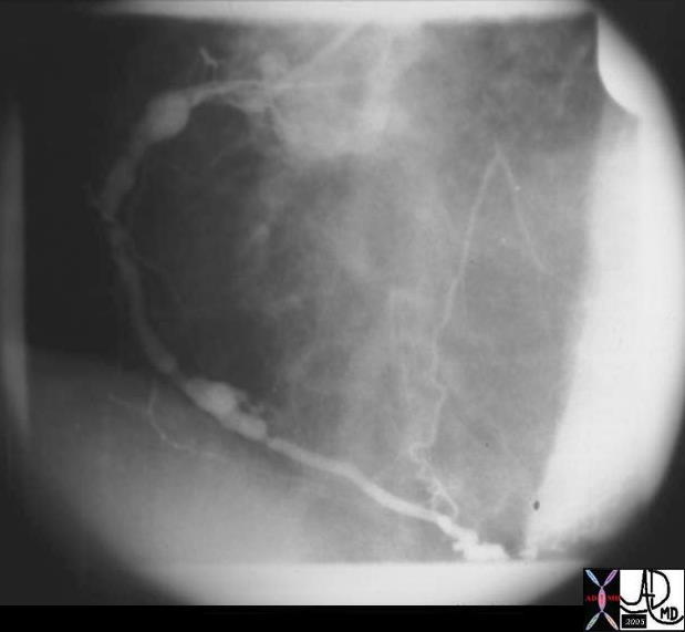
| This angiogram of the right coronary artery shows ectatic and stenotic atherosclerotic disease. Courtsey Ashley Davidoff MD 15384 code heart cardiac coronary artery RCA atherosclerosis atheroma ectasia ectatic A-V nodal coronary AV aneurysm imaging radiology angiography |
Atherosclerotic Aneurysm of LCA |
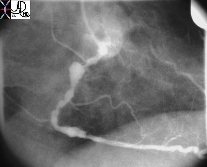
| This angiogram in LAO projection shows stenotic disease and aneurysmal disease of the left coronary artery (LCA)
35354 Courtesy of Laura Feldman MD. code artery atherosclerosis coronary coronary artery heart LCA RCA stenosis atherosclerosis atheroma stenosis circulatory CAD coronary artery disease |
|
Atherosclerotic Aneurysm of LCA |
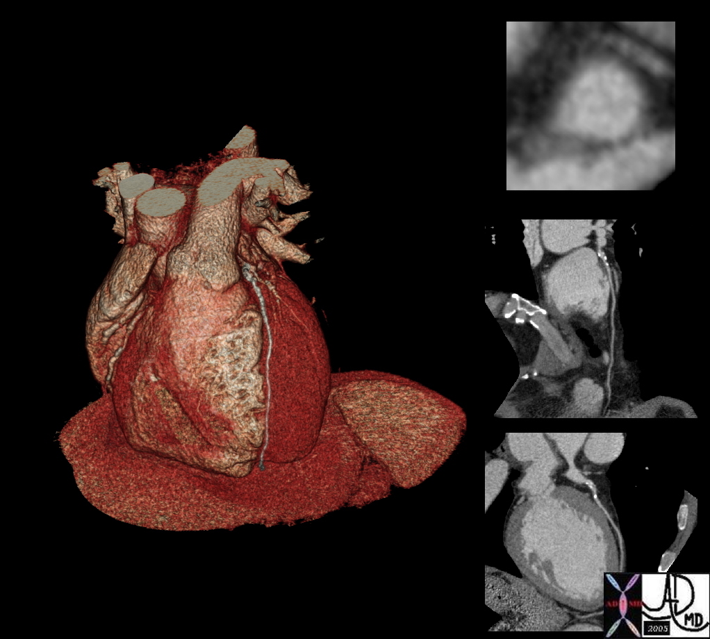
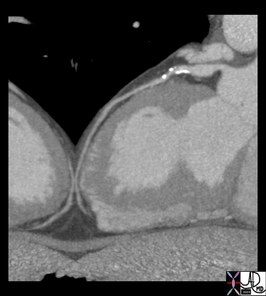
| 44222 heart cardiac artery coronary artery LAD left anterior descending artery fx dilated fx aneurysm dx aneurysmal dilatation imaging radiology CTscan Courtesy Ashley Davidoff MD Jeffrey Mendel MD |
|
||
|
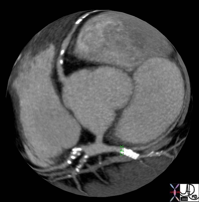
Atherosclerotic Aneurysm ot the origin of the LAD |
| 44250 heart cardiac artery coronary artery LAD left anterior descending artery fx dilated fx aneurysm circumflex fx stent imaging radiology CTscan Courtesy Ashley Davidoff MD Jeffrey Mendel MD |
|
Atherosclerotic Aneurysm ot the origin of the LAD |
| 44254 heart cardiac artery coronary artery LAD left anterior descending artery fx dilated fx aneurysm imaging radiology CTscan Courtesy Ashley Davidoff MD Jeffrey Mendel MD |
|
Atherosclerotic Aneurysm ot the origin of the LAD |
| 44260 heart cardiac artery coronary artery LAD left anterior descending artery fx dilated fx aneurysm circumflex fx stent ramus medianus first diagonal imaging radiology CTscan Courtesy Ashley Davidoff MD Jeffrey Mendel MD |
|
Kawasaki’s Arteritis |
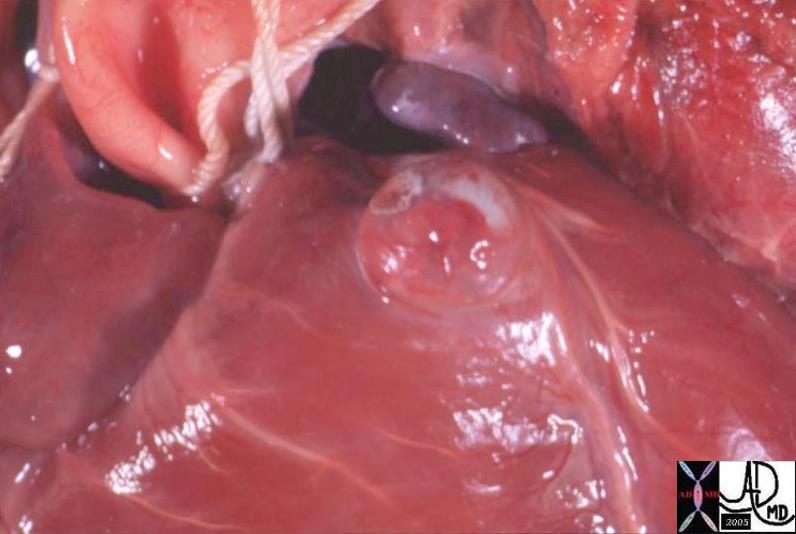
| 07587 heart cardiac aneursym coronary artery Kawasaki’s areteritis grosspathology Courtesy Ashley Davidoff MD |
|
Kawasaki’s Arteritis |
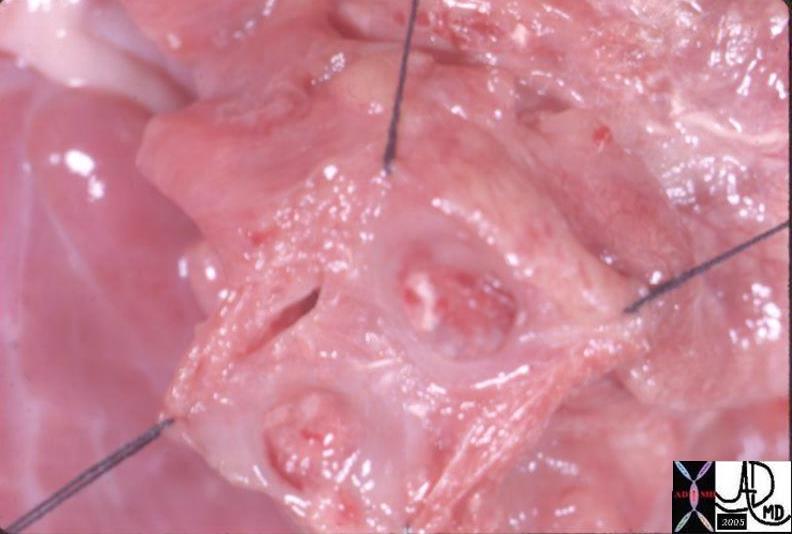
| 07584 heart cardiac aneursym coronary artery Kawasaki’s areteritis grosspathology |

