The Heart
Ashley Davidoff MD

Power of the Heart |
| The artistic rendition of the heart attempts to reveal the ambivalence in the shape of the heartars either a triangular structure or an oval on its side, and it seems to satisfy both shapes in this view. the right ventricle dominates the anterior view and the left ventricle peeks around the left border of the heart holding its power as its trump card behind the right ventricle.
Davidoff art copyright 2009 Courtesy Ashley Davidoff MD 87180b11.8s |

Interior of the RV |
| The artistic rendition of the right atrium and right ventricle. The spatial relationship is best viewed conceptually by tacking the thumb of your right hand and placing it in the middle of the right atrium, and your palm will be aligned with the RVOT code heart cardiac anatomy normal SVC superior vena cava IVC inferior vena cava RA right atrium tricuspid valve right ventricle RVOT right ventricular outflow tract infundibulum conal septum moderator band pulmonary valve
Davidoff art Courtesy Ashley Davidoff copyright 2009 all rights reserved 06566b12b13.4kb05i.8s |

Pulmonary Circulation |
| 32807b09b01.8s heart cardiac pulmonarycirculation pulmonary artery pulmonary vein left atrium Davidoff art copyright 2009 |
|
Artistic rendition of the Left Atrium |
| 77612b.3kb07.8s copyright 2009 all rights reserved Davidoff art |

Bleeding Heart |
| 76329p.8s bleeding hearts nature flowers anatomy copyright 2009 Davidoff photography all rights reserved |
|
The Coronary Tree |
| 15009c.3kb010.8s tree coronaryt tree heart LAD left anterior descending artery Davidoff art Davidoff photography trees in the body copyright 2009 all rights reserved |
|
Chest of Fruit |
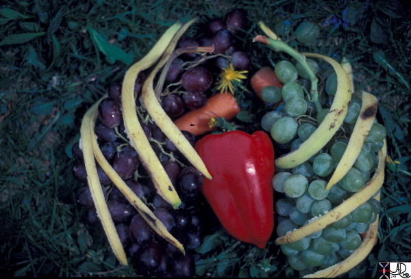
| This artistic rendition of the heart and lungs uses the shape of fruit and vegetables to create an image of the chest. The lungs are made of grapes, the pulmonary arteries are made of carrots, the ribs are made of banana peel and the heat is made of a red pepper. 02032p Courtesy Ashley Davidoff MD. accessory cardiac heart lung bone PA grape banana peel ribs Davidoff art |

Heart in Green |
| The artistic rendition of the heart attempts to reveal the ambivalence in the shape of the heartars either a triangular structure or an oval on its side, and it seems to satisfy both shapes in this view. the right ventricle dominates the anterior view and the left ventricle peeks around the left border of the heart holding its power as its trump card behind the right ventricle.
Davidoff art copyright 2009 Courtesy Ashley Davidoff MD 87180b11b12.8S 87180b11b11.8S |

Scaffolding of the Heart |
| The scaffolding of the heart can be represented by a cross, consisting of two upper smaller components, and two larger lower components. This is only a conceptual 2 dimensional and symmetrical model that will help you understand the structure of the chambers and how the vessels and nerves are arranged around the scaffolding. As the story unfolds the complexity of structure will unfold, but it is important to understand that underlying the complexity there is a simple infrastructure.
Courtesy Ashley Davidoff MD 32058 code cardiac heart introduction infrastructure drawing Davidoff art |
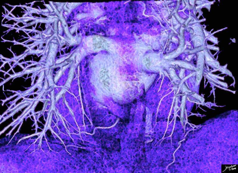
Pulmonary Veins and Left Atrium |
| 77612b.3kb08.8s heart left atrium pulmonary veins red cells normal anatomy copyright 2009 all rights reserved Davidoff art |
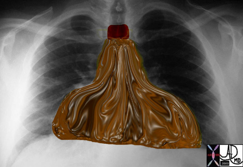
| Water Bottle Heart
|
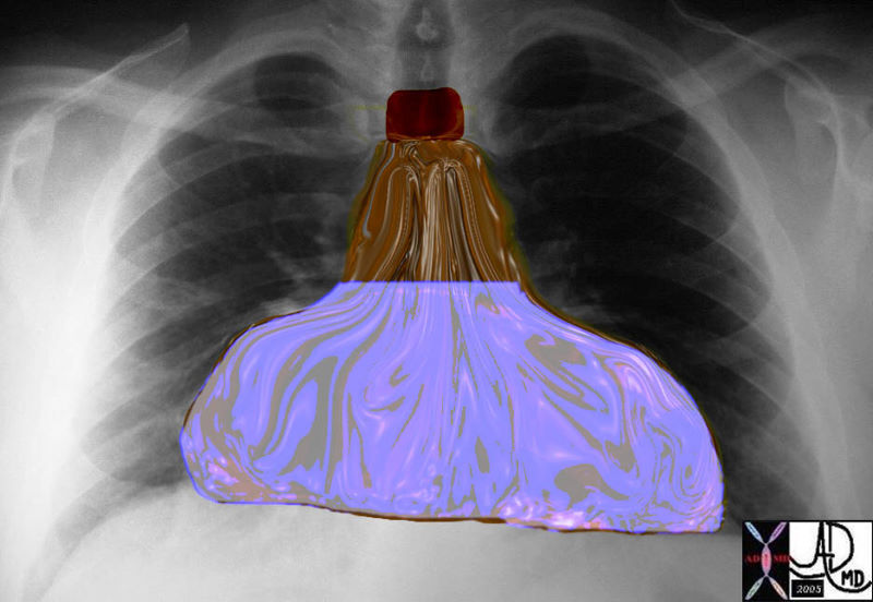
| 30095b08 chest heart pericardium fx waterbottle water bottle dx pericardial effusion Davidoff art |
|
Water Bottle Heart |
| 30095b010 chest heart pericardium fx waterbottle water bottle leather bottle dx pericardial effusion Davidoff art |

Acute MI The Suffering LV Apex |
| 32907b03.61k.8sb060.81s heart cardiac heart myocardium myocyte normal abnormal disease Davidoff Art Copyright 2009 artistic rendition |
|
The Circulation |
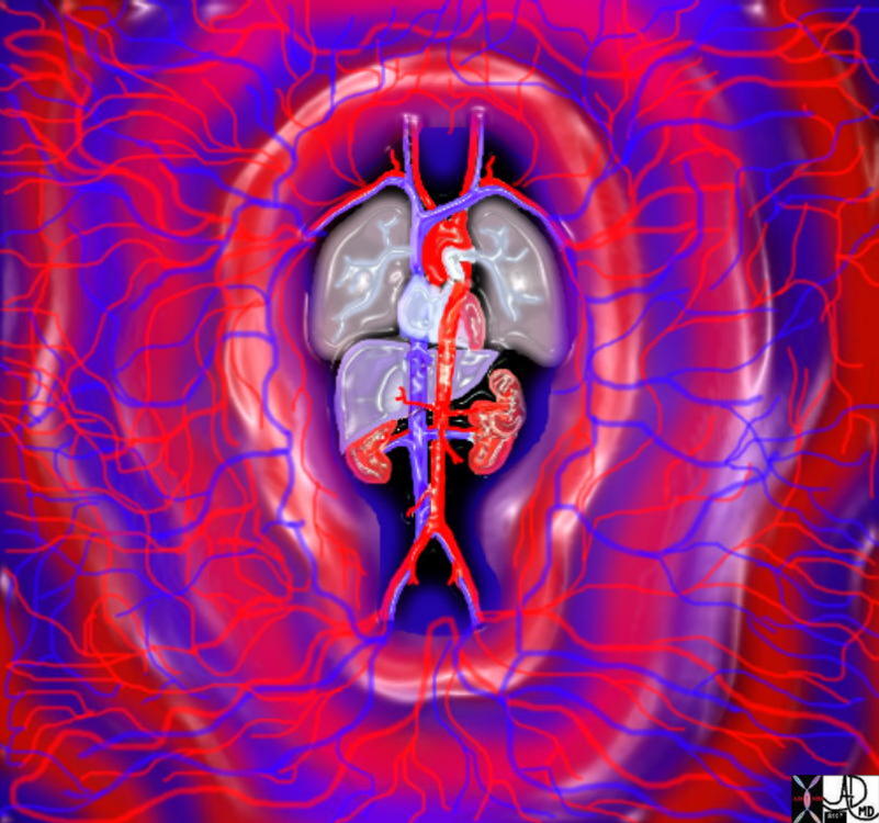
| 01896.800 aorta thorax abdomen thoracic aorta abdominal aorta circulation arterioles venules capillaries normal anatomy physiology pulsatile pulse Davidoff art Courtesy Ashley Davidoff MD |

Valves in Purple |
| 87268b06.33k.8s Cross section through the atrioventricular plane of the heart shows the tricuspid valve (TV), mitral valve (MV) in plane, the aortic valve (AoV) in close association with the mitra valve, and the pulmonary valve (PV). elevated and oriented toward the left The orientation of the coronary arteries in the horizontal plane and their course around the atrioventricular groove is demonstrated. The left main coronary artery arises fromt the left coronary cusp and gives rise to the LAD (left anterior descending artery) and circumflex which travels in the A-V groove around the mitral annulus, and the right coronary artery courses around the tricuspid annulus and gives rise to the PDA code heart cardiac valves mitral valve aortic valve tricuspid valve pulmonary valve MV AoV coronary arteries PDA posterior descending artery LAD left anterior descending artery RCA right coronary artery Cx circumflex TV PV anatomy normal Davidoff art Image modified from Grays anatomy 1918 |
|
Valves in Pink |
| 87268b06.35k.8s 87268c06.8s Cross section through the atrioventricular plane of the heart shows the tricuspid valve (TV), mitral valve (MV) in plane, the aortic valve (AoV) in close association with the mitra valve, and the pulmonary valve (PV). elevated and oriented toward the left The orientation of the coronary arteries in the horizontal plane and their course around the atrioventricular groove is demonstrated. The left main coronary artery arises fromt the left coronary cusp and gives rise to the LAD (left anterior descending artery) and circumflex which travels in the A-V groove around the mitral annulus, and the right coronary artery courses around the tricuspid annulus and gives rise to the PDA code heart cardiac valves mitral valve aortic valve tricuspid valve pulmonary valve MV AoV coronary arteries PDA posterior descending artery LAD left anterior descending artery RCA right coronary artery Cx circumflex TV PV anatomy normal Davidoff art Image modified from Grays anatomy 1918 |
|
The Valves |
| 87268b06.3k.8s Cross section through the atrioventricular plane of the heart shows the tricuspid valve (TV), mitral valve (MV) in plane, the aortic valve (AoV) in close association with the mitra valve, and the pulmonary valve (PV). elevated and oriented toward the left The orientation of the coronary arteries in the horizontal plane and their course around the atrioventricular groove is demonstrated. The left main coronary artery arises fromt the left coronary cusp and gives rise to the LAD (left anterior descending artery) and circumflex which travels in the A-V groove around the mitral annulus, and the right coronary artery courses around the tricuspid annulus and gives rise to the PDA code heart cardiac valves mitral valve aortic valve tricuspid valve pulmonary valve MV AoV coronary arteries PDA posterior descending artery LAD left anterior descending artery RCA right coronary artery Cx circumflex TV PV anatomy normal Davidoff art Image modified from Grays anatomy 1918 |

Carousel Heart |
| 42644.800 lung pulmonary segments parts fissres normal anatomy heart cardiac chambers Davidoff art Davidoff oneness |
|
Aortic Stenosis |
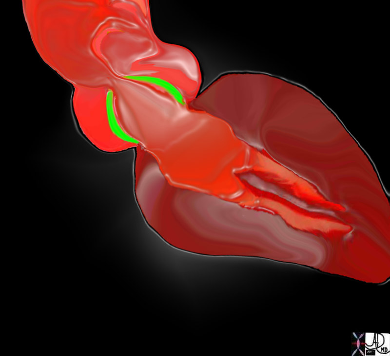
| 07969bW.802 heart cardiac aorta aortic valve fx thickening of the aortic valves LVH left ventricular hypertrophy post stenotic dilatation of the ascending aorta turbulence eccentric jet doming of the aortic valve AV AS aortic stenosis Davidoff art |
|
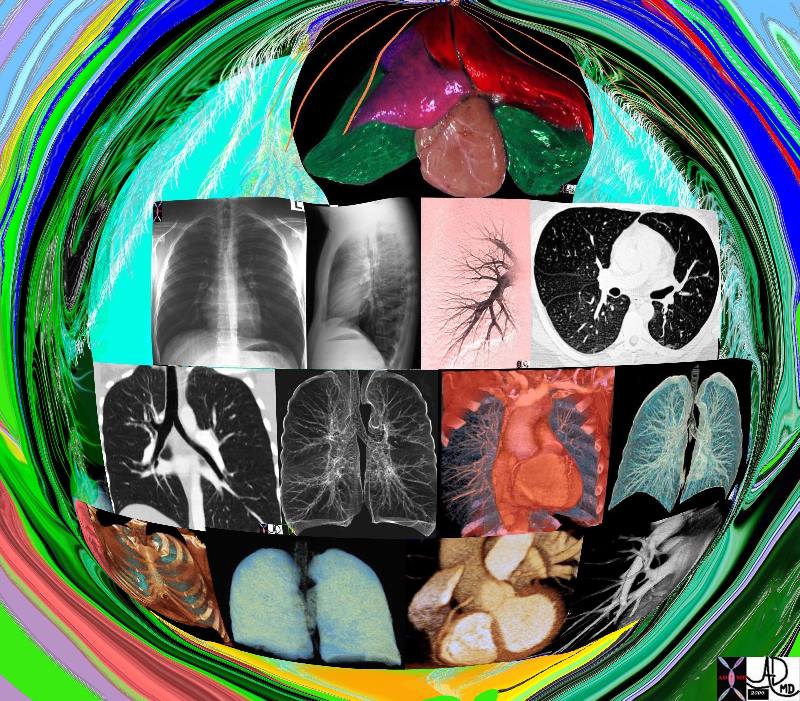
Imaging the Cardiopulmonary Team |
| 42444b17.800 lung chest pulmonary artery bronchus bronchi trachea heart normal anatomy Ddavidoff art Davidoff MD |
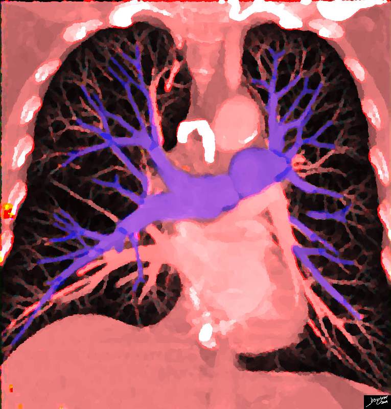
Pulmonary Circulation |
| 32807b03.8s heart cardiac pulmonarycirculation pulmonary artery pulmonary vein left atrium Davidoff art copyright 2009 |

The Normal Heart |
|
The normal heart lies as a gracile structure in the middle of the chest and takes up about 25% of the chest volume. chest lungs fx normal chest CT anatomy Courtesy Ashley Davidoff MD Davidoff art 45774 |

Left Atrial View of the Open Mitral Valve |
| 08407.03.8s left Atrial View of the Open Mitral Valve heart cardiac mitral valve anterior leaflet posterior leaflet MV left atrium LA blood Davidoff art copyright 2009 all rights reserved |
|
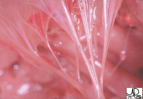
Strings of the Heart |
| Anatomic image of the chordae affixing to the apex of the anterior leaflet of TV. The tip of the anterolateral papillary muscle can be seen at the bottom of the image. This muscle as previously noted is constant and characteristic of the RV. Courtesy of Ashley Davidoff M.D. 32097 |
|
Strings of the Heart From Above |
| The most beautiful feature of the LA (in my opinion of course) is its view of the anterior leaflet of the mitral valve through the annulus. To see the chordae and the sail of the anterior leaflet biphasically flap-flapping must be a sight to behold. Courtesy of Ashley Davidoff M.D. code cardiac heart normal MV mitral valve anatomy 32106 |
|
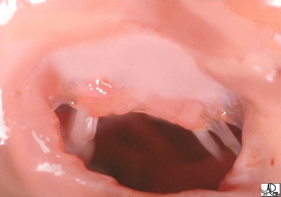
Climb into the Heart and Take a Look at the Mitral Valve from Above |
| If you put your head close to entrance into the
32106.1.5ks04 heart cardiac anatomy normal mitral valve normal anterior leaflet mitral annulus chordae Davidoff photography copyright 2008 pathology high resolution
LV you will be faced with this breath taking view of the anterior leaflet of the MV with its attached chordae.
|
|
Maid of the Mist in the Mitral Valve |
|
Lean a little more forward and the sound you will hear is similar to the sounds of the Maid of the Mist in the middle of Niagara Falls. In addition, you will see how the glistening fibers of the chordae are attached to the papillary muscles of the left ventricle. 32118b03 heart papillary muscles chordae tendinae left ventricle maid of the mist mitral valve sounds of the blood flow Davidoff art Davidoff photography copyright 2008 |
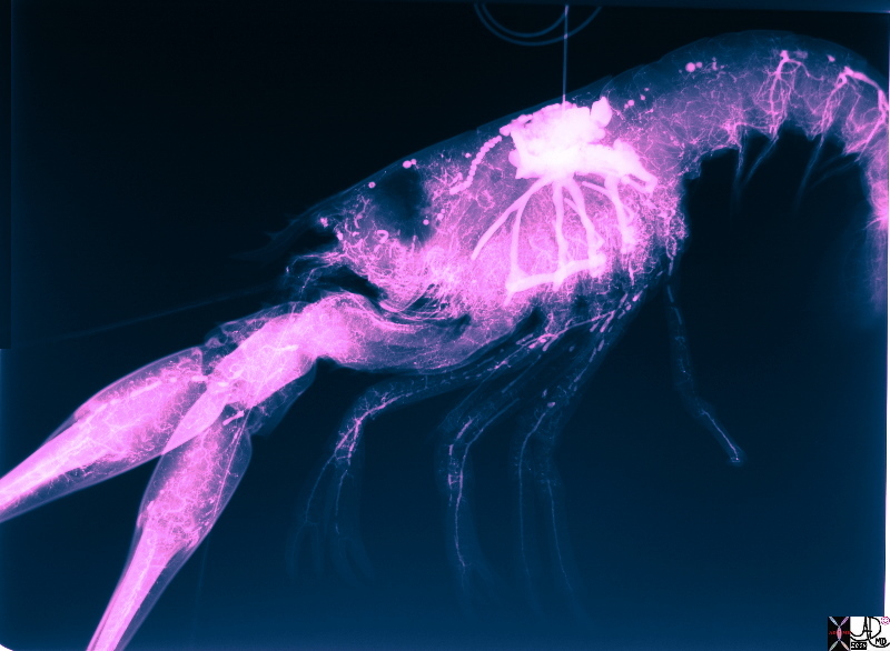
Lobster Circulation |
| The heart of the lobster has been injected with contrast and the circulation filled with contrast revealing head to toe vasculature
Courtesy Dr Deborah Termeulen copyright 2009 49790b01.8s |


