Copyright 2009
Left Heart Failure
-
- Heart failure due to left ventricular dysfunction
- heart failure with reduced ejection fraction (HFrEF)
- ejection fraction (EF) of less than 40%.,
- heart failure with preserved ejection fraction (HFpEF), and
- heart failure with an EF of greater than 50%.
- heart failure with mid-range ejection fraction (HFmrEF).
- EF of 40% to 50% T
- heart failure with reduced ejection fraction (HFrEF)
- Heart failure due to left ventricular dysfunction
|
Acute Pulmonary Edema |
| In this patient with acute congestive cardiac failure the consolidation that has hilar distribution has reminded radiologists of bat wings and is caused by alveolar edema. As a result of the fluid in the alveoli, gas exchange across the respiratory membrane is reduced and required intubation to improve the gas exchange process. Note the endotracheal tube as well as the central venous line that is used to assess the heart pressure and monitor the congestion. Courtesy Ashley Davidoff MD 42073b01 heart cardiac interstitia; edema alveolar edema batwing CHF congestive cardiac failure CXR plain film |
|
Normal Alveoli |
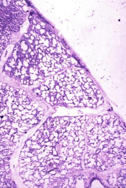
| Pleura Note thin mesothelial membrane lying on top of alveolated lung parenchyma. This represents the visceral pleura. Courtesy Armando Fraire MD 32648 code lung pulmonary pleura normal alveolus alveoli histology interstium interstitial |
|
Normal Secondary Lobule Interlobular Septa |
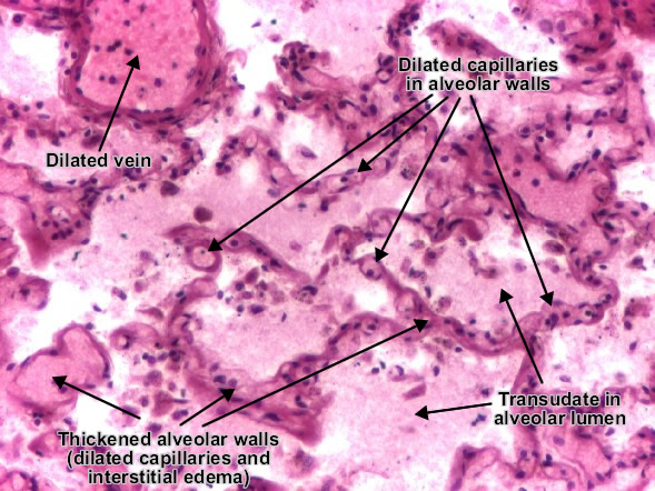
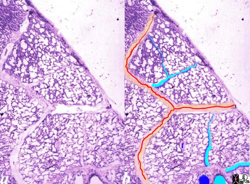
| Normal lung histology This image is a panoramic view of the lung showing secondary lobules and interlobular septa. Within the interalveolar septae, one sees small venules and lymphatics.Courtesy Armando Fraire MD. 32649b code lung pulmonary alveoli alveolus secondary lobule interlobular septa vein lymphatic histology interstitium interstitial |
|
Normal Secondary Lobule |
| The arteries and airways pair up and travel together down the respiratory tree branching in exactly the same way until they reach the pulmonary lobule. The pulmonary lobule, also called the secondary lobule is a structural unit surrounded by a membrane of connective tissue, and it is smaller than a subsegment of lung but larger than an acinus. This diagram shows two secondary lobules lying side by side. The pulmonary arteriole (royal blue) and bronchiole (teal) are shown together in the centre of the lobule (“centrilobular”), with two other pairs of bronchovascular bundles, while the oxygenated pulmonary venules (red) and lymphatics (yellow) are peripheral and also form formidable and almost inseparable pairs.
Courtesy Ashley Davidoff MD Copyright 2009 42440b03 |
|
Cephalization |
| 44659.800 chest lung CXR cephalisation cephalization pulmonary arteries pulmonary artery fx distended distension fx pulmonary congestion dx CHF dx congestive heart failure cardiac imaging radiology Courtesy Ashley Davidoff MD |
|
Size of the Upper Lobe Brancjh artery to airway Ratio |
| 44659c03.800 Copyright 2009 Davidoff MD |
|
Interstitial Edema |
| 46421 heart cardiac chest lung fx pulmonary congestion bilateral effusions interstitial edema Kerley B lines thickening of the fissure left atrial enlargement elevation of the mainstem bronchus cardiomegaly ‘fx enlarged dx congestive heart failure CHF cardiac failure CXR plain film Davidoff MD 46424c01 46421 46422 46423 46424 |
|
Kerley B Lines |
| 46423 heart cardiac lung chest fx pulmonary congestion interstitial edema Kerley B lines Kerley A line Kerley C lines dx congestive heart failure CHF cardiac failure CXR plain film Davidoff MD 46424c01 46421 46422 46423 46424 |
|
Progression From Normal to Complete White Out |
| 41972c Courtesy Ashley Davidoff MD medical students code alveolar edema cardiac cardiac failure congestion enlargement heart lung whiteout |

87 year old male with mitral stenosis and mild MR with enlarged left atrium, and right ventricle with pulmonary hypertension and probable mild LV enlargement as well
Ashley Davidoff MD

Ashley Davidoff MD
Keywords:
lung pulmonary alveoli alveolus secondary lobule interlobular septa vein lymphatic histology interstitium interstitial normal copyright 2020 all rights reserved
Courtesy of: Armando Fraire, M.D.
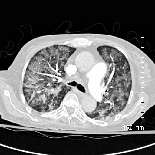
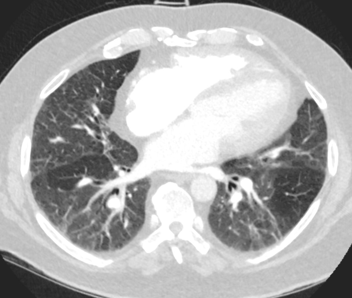
CT scan shows Diffuse ground glass pattern with thickening of the interlobular septa and manifesting as crazy paving pattern
Ashley Davidoff MD
References and Links
Malik, A, et al Congestive Heart Failure Stat Pearls September 2021
Ahmad;
-
Links from TCV
- Intro
- Diastolic Heart Failure
- Right Heart Failure
- Pulmonary Edema
- CXR
- CT
- Cases
- 003H Non Compaction CHF and Pacemaker
- 020H 34M CRF Dilated Cardiomyopathy Diabetes Pancreatitis
- 024H CHF Kerley b CXR CT
- 025H Sickle Cell CHF Spleen Bone
040 Sickle Cell and CHF
042 CHF Kerley B and A - 047H Acute MI Left Main Occlusion Batwing CXR
- 049H Early CHF CXR Equalization
- 049Hb CHF Cephalization in progress
- 050H CHF, CXR, and Interstitial Edema in progress
- 072H 49M CAD CHF ground glass


