Conotruncus Unknown Cases
At the level of the
Ao-PA
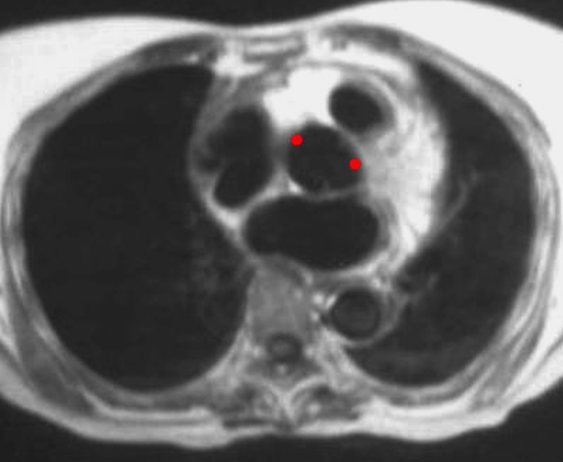
Normal Relationship of the pulmonary Artery to the Aorta
Ashley Davidoff MD The CommonVein.net
Understanding
The conotruncal abnormalities (including, Tetralogy of Fallot, transposition, truncus arteriosus, double outlet right ventricle and double outlet left ventricle require understanding of the embryogenesis of the heart
“Nothing in life is to be feared, it is only to be understood. Now is the time to understand more, so that we may fear less.”
― Marie Curie
The noblest pleasure is the joy of understanding.

Albert Einstein

Sizes of the Cardiovascular System
Embryology
From a straight tube to complex structure
1- Looping of the Heart
2- Septation
3- Connecting the Parts
1- Looping
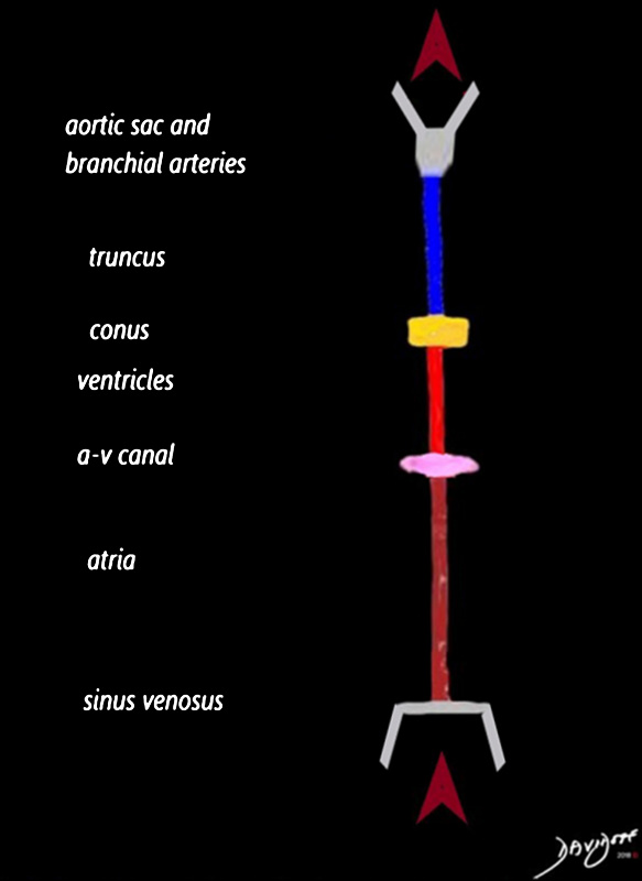
Blood enters the sinus venosus and into the developing atria (maroon), after which it traverses the region of the developing A-V canal (pink) and into the developing ventricles (red) The blood passes through the conus (yellow) and via the truncus (blue)into the aortic sac and branchial arch arteries
Ashley Davidoff MD
TheCommonVein.net

Ashley Davidoff MD
TheCommonVein.net
RV goes Right and Anterior in the D Loop
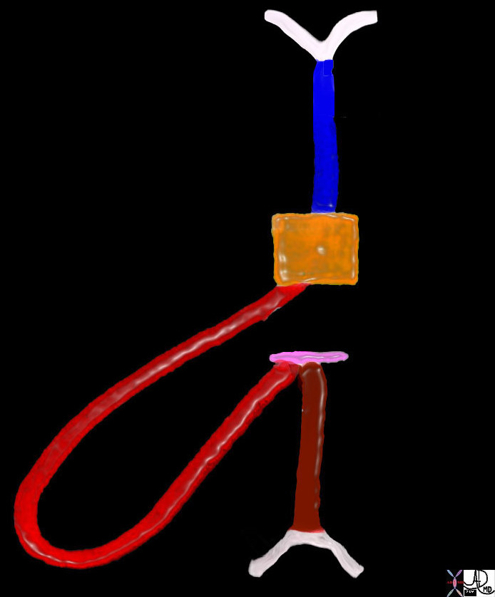
The developing RV in most hearts starts out by looping to the right and also evolves anteriorly because there is room anteriorly in the chest for development since the tube starts out abutting the spine
keywords heart cardiac straight tube d loop D loop primitive heart embryology right ventricle RV sinus venosus atria atrium A-V cushions conus arteriosus truncus arteriosus drawing Ashley Davidoff Davidoff MD 01488b02
LV arises as a daughter ventricle from the RV and goes left and posterior with atria even further posterior
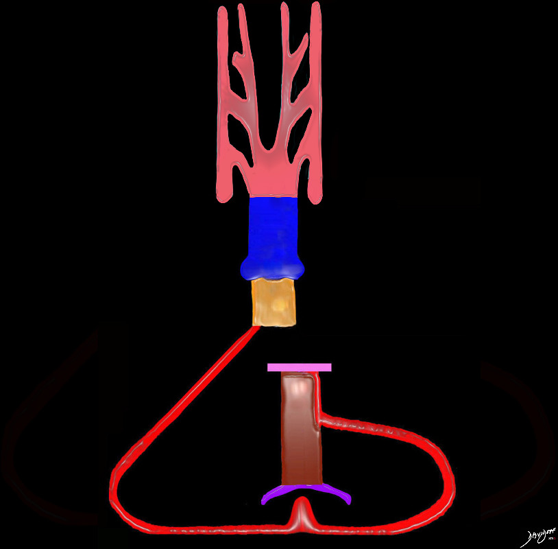
The left ventricle develops from the right ventricle but lies posteriorly. At this stage The heart prior to septation both ventricles have their outlet through the right ventricle prior to septation- (ie double outlet right ventricle phase ), so blood flows into the common ventricular chamber, and out into the conus (infundibulum) and into the truncus prior to entering the aortic sac
Ashley Davidoff MD
TheCommonVein.net
DORV Morphology and Physiology – One Big Mix of Blood
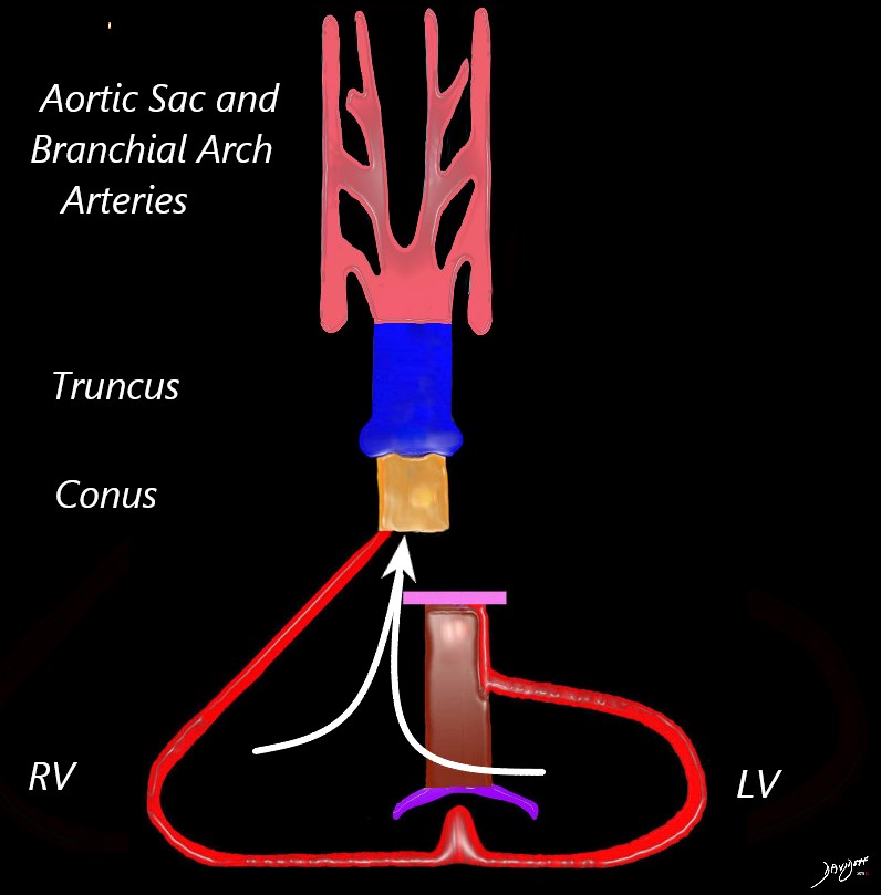
The heart in double outlet right ventricle phase prior to septation, so blood flows intot the common ventricular chamber, and out into the conus (infundibulum) and into the truncus prior to entering the aortic sac
Ashley Davidoff MD
TheCommonVein.net
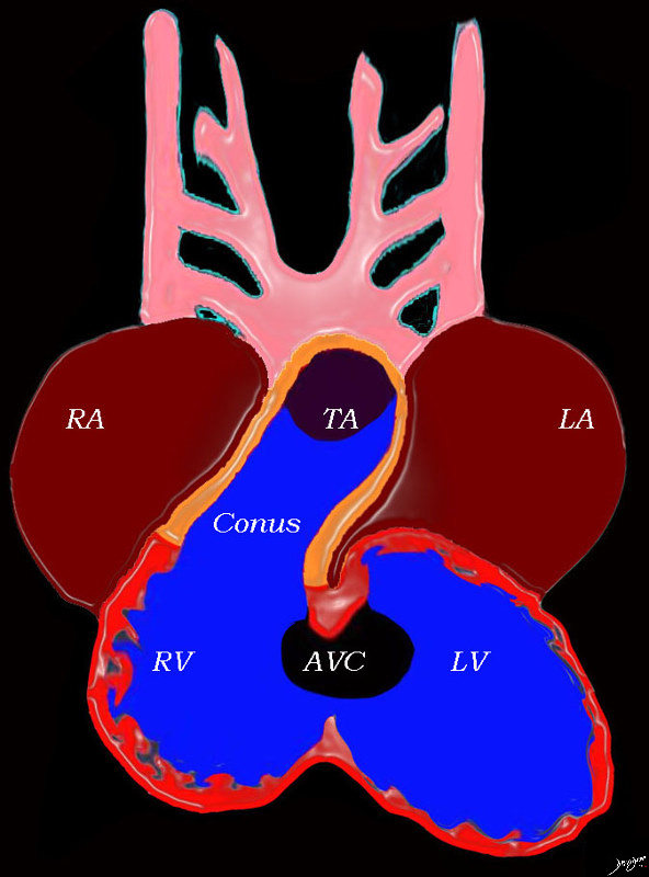
At this stage the heart consists of un-septated atria, ventricles and great vessels, with atria situated posteriorly connected to the ventricles by the AV canal, and the ventricles have a common outlet over the right ventricle to the conus arteriosus, which is connected to the truncus arteriosus
Key words heart cardiac right atrium left atrium right ventricle, left ventricle atrioventricular canal conus arteriosus truncus arteriosus aortic arches RA LA RV LV AVC embryology 01492b04L Ashley Davidoff MD TheCommonVein.net
DORV Phase

The heart in double outlet right ventricle phase prior to septation, so blood flows intot the common ventricular chamber, and out into the conus (infundibulum) and into the truncus prior to entering the aortic sac
Ashley Davidoff MD
TheCommonVein.net
Focus on the Conotruncus
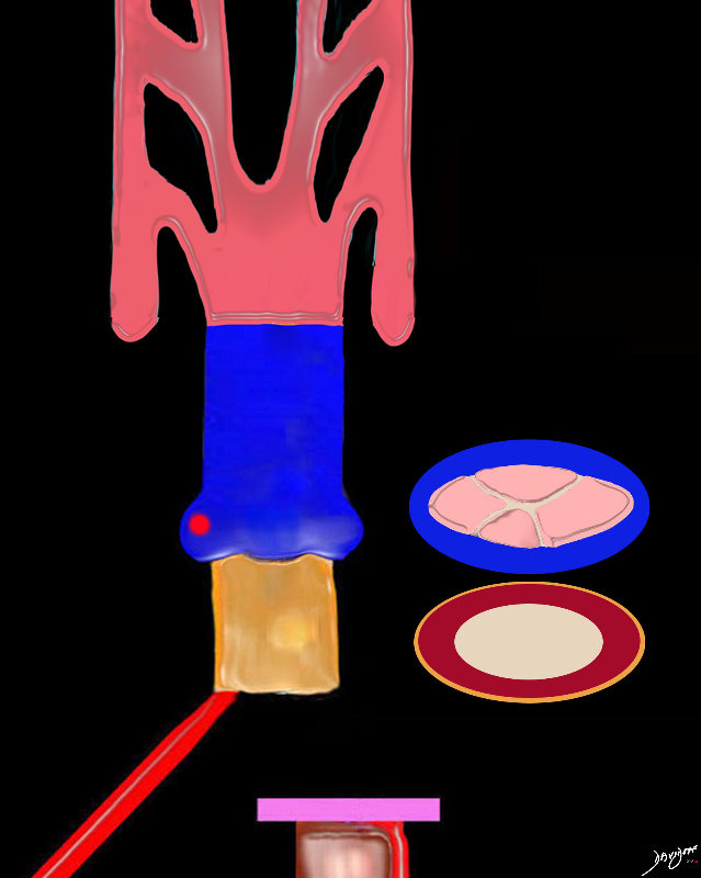
The conus (gold exterior) consists of a single muscular tube prior to septation.
The origin of the truncus contains 4 truncal cushions which are going to develop into the aortic and pulmonary valves following septation.
Ashley Davidoff MD
TheCommonVein.net
2 -Septation of the Conotruncus
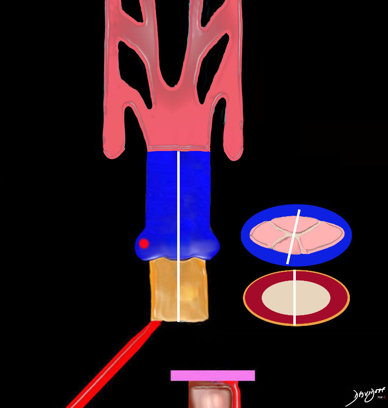
A D loop will result in positioning the aorta to the right of the pulmonary artery, but at this stage the conus still arises from the developing right ventricle (DORV).
The truncus after it divides will have two valves consisting of 3 leaflets each
During septation the conus will be divided into a right side portion that will be subaortic, and a left sided portion that will be subpulmonary
Ashley Davidoff MD
TheCommonVein.net
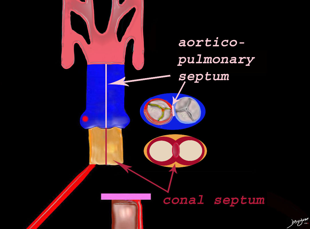
The conus consists of muscle bound tubes, separated by the conal septum, and the aorta and pulmonary artery are separated by the aorticopulmonary septum. The morphology and physiology are consistent with a double outlet right ventricle state. Both great vessels receive mixed blood from the “single” ventricle.
Ashley Davidoff MD
TheCommonVein.net
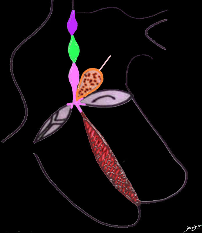
The atrial septum consists of 3 parts; The most superior is the septum derived from the sinus venosus (purple), the middle is the septum primum (green) and the most inferior is derived from the endocardial cushions (pink) The endocardial cushions also contribute to the formation of the of the membranous component of the interventricular septum. The interventricular septum consists of the membranous septum and the muscular septum (red). The great vessels are separated by a muscular conal septum (orange) and the membranous aorticopulmonary septum (light pink).
Ashley Davidoff MD TheCommonVein.net

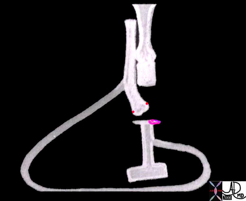
At this stage the heart is close to totally septating, but before this happens the left ventricle and aorta have to connect in order to create a systemic circulation that is separate from the pulmonary circulation. The aorta lies to right of the pulmonary artery and still has a subaortic conus. In order to connect with the left ventricle and mitral valve, the subaortic conus has to resorb.
Ashley Davidoff MD TheCommonVein.net
3- Connecting the Parts

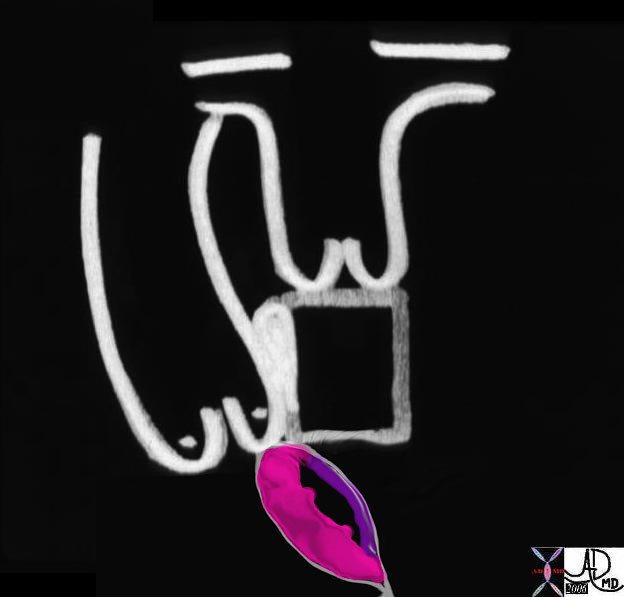
At this stage the heart is close to totally septating but before this happens the left ventricle and aorta have to connect in order to create a systemic circulation that is separate from the pulmonary circulation. The aorta lies to right of the pulmonary artery and still has a subaortic conus. In order to connect with the left ventricle and mitral valve, the subaortic conus has to resorb. Mr Aorta has a his eyes set on Ms Mitral Valve
Ashley Davidoff MD TheCommonVein.net
A Poem –
A Marriage Made in Heaven
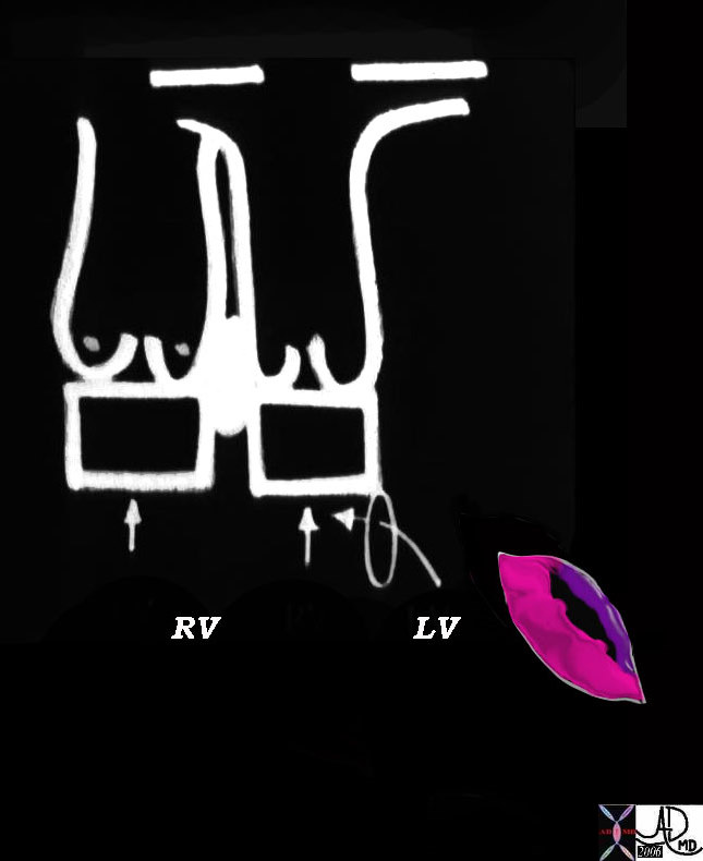
Mr Aorta only has eyes for Ms Mitral
How is Mr Aorta going to reach her?
Double Outlet RV Morphology
Ashley Davidoff MD
07427b10
TheCommonVein.net
keywords
06370b04 heart cardiac bilateral conus outflow tract infundibulum aorta pulmonary artery D-loop RV LV cono-ventricular defect bilateral conus atrioventricular endocardial cushion mitral valve anatomy embryology subaortic conus sub-pulmonary conus fibrous continuity mitral valve MV
06370b04
Ashley Davidoff MD
TheCommonVein.net
07427b02
Ashley Davidoff MD
TheCommonVein.net
A Poem A Marriage Made in Heaven
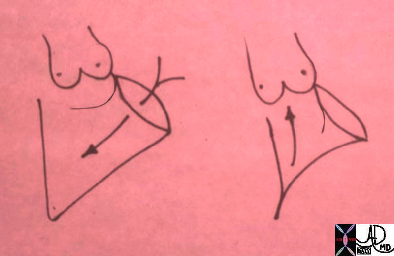
The mitral valve participates in the inflow as well as the outflow of the LV.
The LV does not have an infundibular chamber like the RV.
It is the anterior leaflet of the MV that has this dual function.
The first drawing represents LV diastole, showing the open anterior leaflet acting as the anterior, medial and rightward border of the inflow to the LV.
The second drawing is the systolic phase where this same anterior leaflet acts as the leftward and lateral border of the outflow tract.
Ashley Davidoff MD
TheCommonVein.net
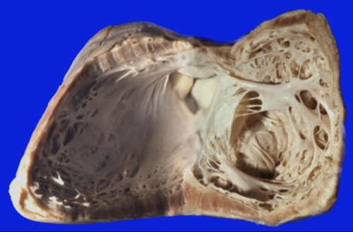
In this post mortem image the left ventricle is opened along the LAD anteriorly and PDA posteriorly so that the ventricular septum is displayed to the left and the free wall, mitral valve and papillary muscles to the right. The aortic valve is seen to the top of the image in fibrous continuity with the anterior leaflet of the mitral valve. Note the conical shape of the left ventricle, the relatively smooth septal wall that has fine muscle bundles, the two major groups of papillary muscles, and the relatively thick muscular wall.
key words
heart LV left ventricle mitral valve MV ventricular septum free wall anterolateral papillary muscle posteromedial papillary muscle aortic valve fibrous continuity normal anatomy gross anatomy
Ashley Davidoff MD
15389b01
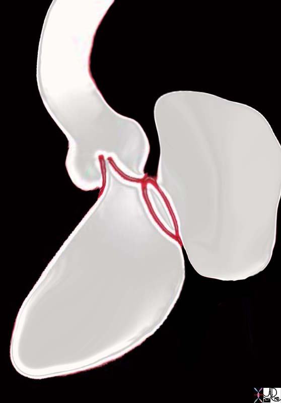
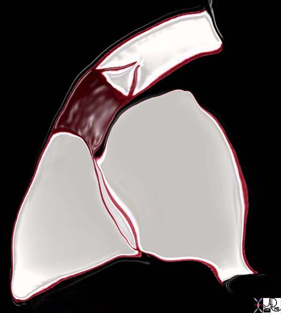
Note how the aorta is tucked facingposteriorly (“heels of the patient”) as a result of the conal resorption
06373b05 heart cardiac aorta mitral valve anatomy embryology resorption of subaortic conus fibrous continuity mitral valve with aortic valve position shape of aorta MV Ashley Davidoff
TheCommonVein.net
key words
heart cardiac pulmonary artery growth of the subpulmonary conus pulmonary valve infundibulum RV right ventricle RA right atrium IVC inferior vena cava tricuspid valve anatomy embryology position shape of pulmonary artery
Courtesy Ashley Davidoff Davidoff drawing 06376b04 TheCommonVein.net
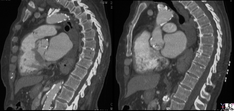
Keywords infundibulum right ventricle RVOT right ventricular outflow tract aortic valve aortic sclerosis calcification enlarged left atrium heart normal conotruncal relationship cardiac Courtesy Ashley Davidoff MD
31149c.8
Ashley Davidoff MD
TheCommonVein.net
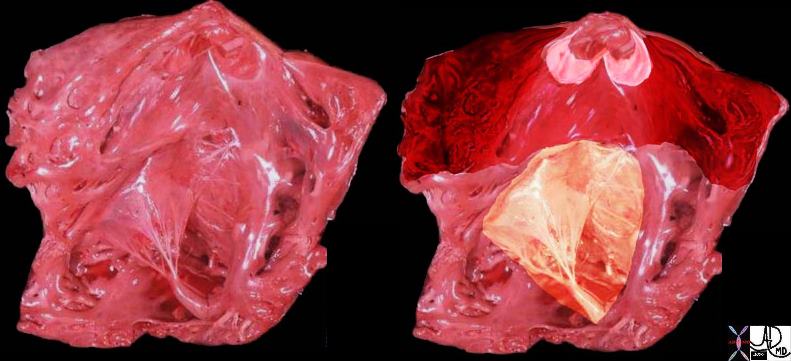
The tricuspid valve is overlaid in yellow showing the large anterior leaflet, and medially placed septal leaflet.
cardiac heart right ventricle RVOT right ventricular outflow tract parietal band septal band conal septum papillary muscle of Lancisi conal papillary muscle anterolateral papillary muscle pulmonary valve pulmonary artery ventricular septum trabeculae carneae normal anatomy gross anatomy Davidoff MD 06409c01
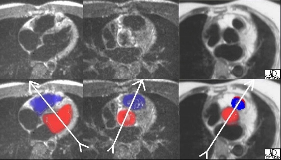
The “twist” story is graphically demonstrated in the following axial MRI images, cutting from inferior to superior as we follow the RV sinus, and RVOT around the aorta. Image 1 shows the triangular RV sinus anterior and to the right of the left ventricle. Image 2 is more superior and shows the RVOT making its way leftward and anterior to the aorta. Image 3 shows the final, distal, and leftward position of the RVOT just below the pulmonary valve. The next set of images with color overlay may be helpful. Ashley Davidoff M.D. 32093 TheCommonVein.net

embryology bilateral conus DORV growth resorption normal mitral to aortic continuity transposition D transposition double outlet right ventricle
Ashley Davidoff art copyright 2019
06394c01L.800s
TheCommonVein.net
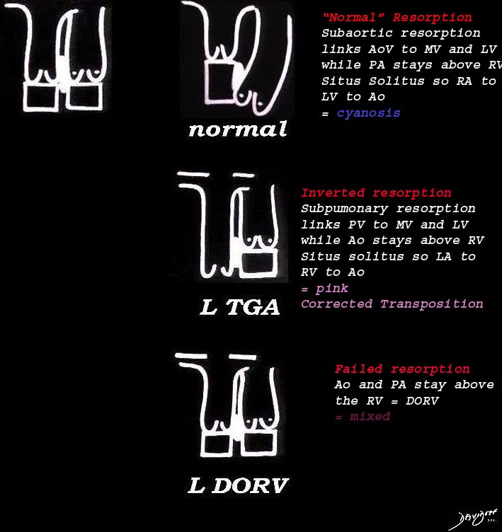
embryology bilateral conus DORV growth resorption normal mitral to aortic continuity transposition D transposition double outlet right ventricle
Ashley Davidoff
06394c02L01s
TheCommonVein.net
Anatomy of the RV

Tricuspid Valve and Pulmonary Valve
The first diagram is a simple drawing of the right ventricle as seen in a frontal projection. The tricuspid valve is to the right (patient position)and inferior and the pulmonary valve to the left and superior. The arrows of the second diagram show the inflow portion of the right ventricle and the outflow portion. The third and last diagram shows the two chambers that make up the right ventricle. The right ventricular inflow chamber also called the RV sinus is triangular and in orange while the outflow chamber is more tubular or cylindrical and has been called many names – but somehow it does not seem to care. Right ventricular outflow tract (RVOT), and infundibulum seem to be the most popular. Courtesy of Ashley Davidoff M.D. 32087 06610 b trio
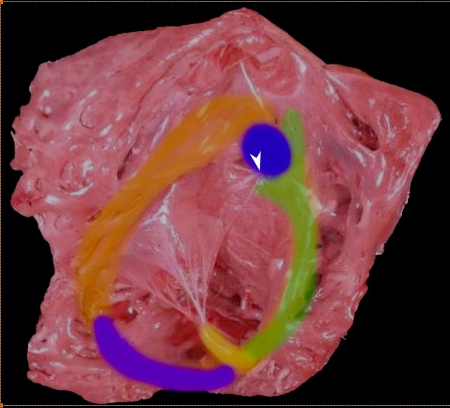
Thr superior aspect of the septal band (green) has two limbs – the “Y” of the septal band. and the conal septum (blue) embeds itself in the Y of the septal band. The (chordae) of the conal papillary muscle (aka papillary muscle of Lancisi- white arrow) also inserts in the “Y ” of the septal band. The anterolateral papillary muscle (yellow) )originates from the septal band) and subtends the anterior leaflet of the tricuspid valve . The moderator band (purple) connects the septum to the the free wall, and the parietal band (orange) completes the muscular ring that separates the inflow from the outflow tract. The pulmonary valve (above the parietal band and conal septum) defines the fborder between the RVOT and conus with the pulmonary artery.
Ashley Davidoff MD
06409 RV muscle bands b01.81

This axial MRI view shows the RVOT anterior and leftward of the aortic valve, after it has twisted around the aorta. The thin musculature of the ovoid infundibulum is in red overlay in the second image.
Ashley Davidoff MD
TheCommonVein.net
07523e.81sL
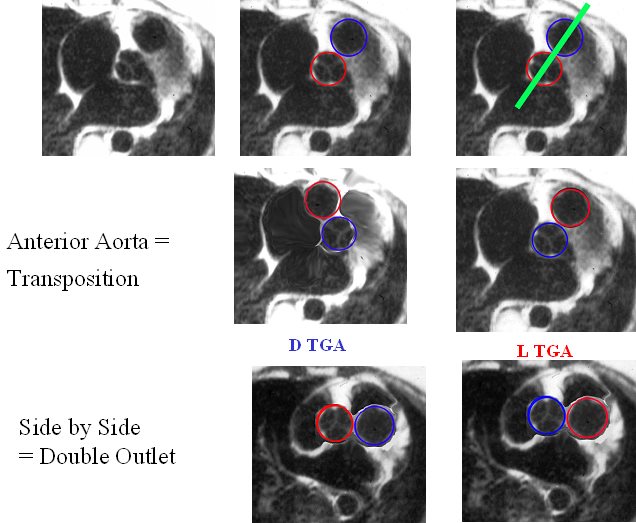
The image reflects the relationship of the aorta and pulmonary arteries in the normal patient, in DTGA, LTGA and DORV. In the normal patient with D loop the aorta (Ao) is posterior and to the right, and the pulmonary artery (PA) is anterior and to the right. In the patient with DTGA, the Ao is anterior and to the right and the PA is posterior and to the left. In an L loop the Ao is anterior and to the left and the PA is posterior and to the right. In double outlet right ventricle (DORV) the great vessels lie side by side and in DORV with a D loop the aorta is to the right and with an L loop the aorta is to the left
86778 01639 01639d02 01639d04 01639f03.jpg Anterior aorta = Transposition
Ashley Davidoff MD
TheCommonVein.net
References
Restivo A et al Cardiac outflow tract: A review of some embryogenetic aspects of the conotruncal region of the heart The Anatomical Record First published: 04 August 2006 Radiographics
Luba et al Cardiovascular MR Imaging of Conotruncal Anomalies Radiographics
- TCV
- Conotruncal Abnormalities
- Embryology
- Anatomy of the Right Ventricle
- Double Outlet Right Ventricle
- Double Outlet Left Ventricle
- Tetralogy of Fallot
- Transposition of the Great Vessels
- Truncus Arteriosus
