
Lessons of the Past
CXR
Heart Frontal View
Frontal View Applied to Abnormalities
Lateral View Basic Parts Lateral View
CXR and CHF
CT- MRI
Normal size of the chambers am wall on CT and MRI
Sarcoid most common LGE is subepicardial (superficial)
Amyloid most common LGE subendocardial spares apex
Ischemia Subendocardial
The Coronary Arteries
A Little Art – A Lot of Coronary Artery PowerPoint
Etymology “Corona” = Crown or Wreath

Corona = Crown or wreath from mid 17th century (in the sense ‘resembling a crown’): from Latin coronarius, from corona ‘wreath, crown’.
Courtesy Ashley Davidoff MD
Corona Virus – Same etymology
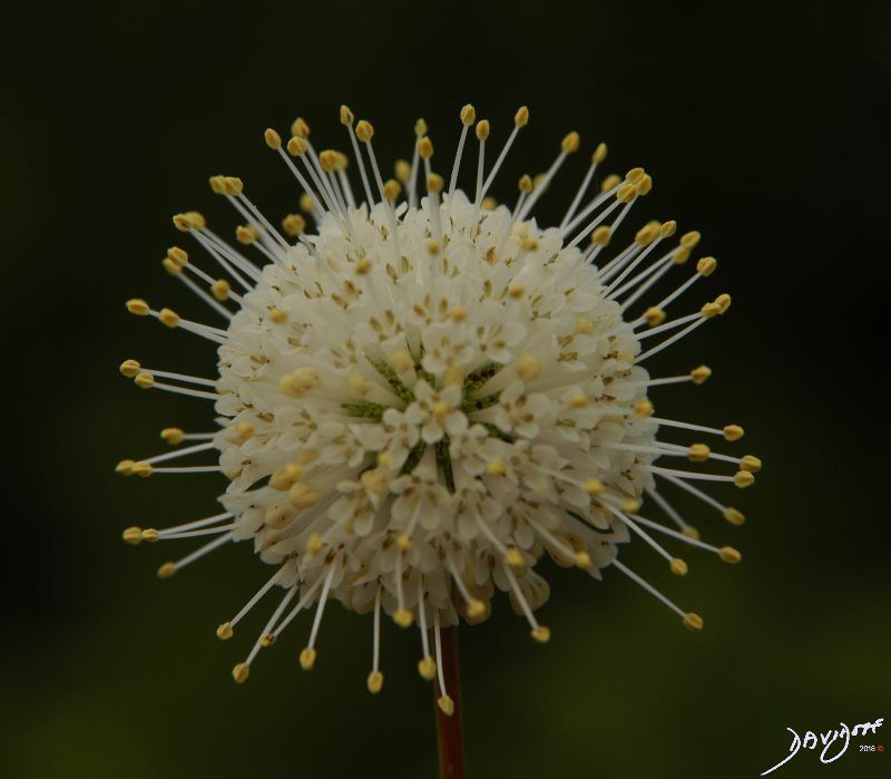 The shape of the corona virus causing COVID-19 bears similarity with the flower of the Buttonbush (Cephalanthus) pictured above The common buttonbush blooms in June through September and sets fruit in September and October. Key characteristics of common buttonbush are its pincushion flower heads, elongated lenticels, and swollen stem base
Interesting Features About the Coronary Arteries
- Supplies its own Circulation
- The heart is the only organ that supplies its own circulation.
- Myocardial oxygen consumption vs Skeletal Muscle
- In humans, at a heart rate of 60–70 beats/min the oxygen consumption normalized per gram of myocardium is 20-fold higher than that of skeletal muscle at rest. As an adaptation to this high oxygen demand, the heart maintains a very high level of oxygen extraction of 70–80% compared with 30–40% in skeletal muscle.2
- Capillary density vs Skeletal Muscle
- This is facilitated by the capillary density of 3000–4000/mm2, compared to 500–2000 capillaries/mm2 in skeletal muscle, and a tight regulation of the coronary blood flow.3
- Capillary density is less in the interventricular septum and AV nodal tissue making them more susceptible to ischemia, putting the conduction systems in these areas at a relatively higher risk to ischemia
- Hilum?
- The heart does not have a hilum where blood flow enters and leaves and receives its blood flow from the periphery and venous drainage go from the center to the periphery The most vulnerable part of the heart therefore for ischemia is the subendocardial region and hence for cardiac MRI the almost pathognomonic Infarction an ischemia therefore
The Heart, The Cross, and The Crux of the Heart
Infrastructure or Scaffolding of the Heart

The Basilica of the National Vow is a Roman Catholic church located in the historic center of Quito, Ecuador. It is the largest neo-Gothic basilica in the Americas
The devotion to the Sacred Heart (also known as the Most Sacred Heart of Jesus is one of the most widely practiced and well-known Catholic devotions The heart of Jesus is viewed as a symbol of “God’s boundless and passionate love for mankind”
Key words heart, cross sacred heart, Basilica, Quito Ecuador
Ashley Davidoff MD

The infrastructure of the heart is a cross resulting conceptually in a box with four chambers. The upper chambers are the receiving chambers and the lower chambers are the pumping chambers. Between the two atria is the interatrial septum, and between the two ventricles is the interventricular septum.
This diagram shows perfect symmetry. The heart although having the same infrastructure has a beautiful asymmetric symmetry, similar to the painting of a Girl Before the Mirror by Picasso.
Ashley Davidoff MD
Relative Position of the RCA vs the LCA and Position of the Right Sided Structures compared to the Left
A Mike Tyson Upper Cut with the Right

Right Sided Structures Right Sided, Anterior and Inferior, Except the Pulmonary Artery which is Leftward and Superior
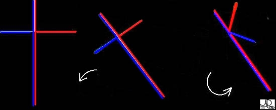
The cross lies with right side inferior, and rotated clockwise so the right sided structures are inferior and anterior and the left sided structures are superior leftward and posterior
Ashley Davidoff MD
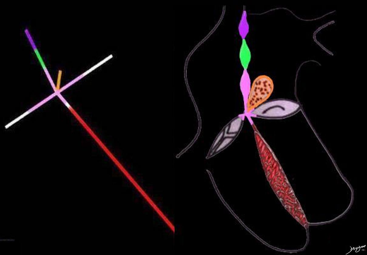
This diagram is slightly more complex but in general reveals the same concepts as described above, and shows the position of the right sided tricuspid valve and the mitral valve. In this diagram the crux of the heart and the endocardial portion of the inter-atrial septum and inter-ventricular septum are colored in pink. The inter-atrial septum consists of the sinus venosus component (purple), the septum secundum (green) and the septum primum (pink) The inter-ventricular septum consists of a muscular portion (red), and a endocardial cushion component (pink). The conal septum is part of the conus arteriosus, lies between the pulmonary artery and aorta and is colored in orange.
Ashley Davidoff, M.D.
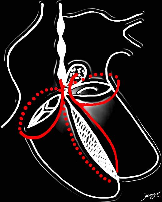
The heart has a left and right arterial system. The vessels are formed around the cross of the heart, “vertically” along the inter-ventricular septum and inter-atrial septum, and “horizontally” in the atrioventricular (A-V) grooves. The left coronary artery arises from the left coronary ostium and supplies one branch, the left anterior descending artery, that travels anteriorly on the anterior aspect of the interventricular septum, and one branch that travels in the atrioventricular groove (left circumflex). The right coronary artery has a branch that courses along the right atrioventricular groove and usually continues as the posterior descending artery on the posterior aspect of the interventricular groove, and commonly gives rise to the A-V nodal branch, at the back of the crux of the heart, which travels in the vertical axis in the interatrial septum.
Courtesy of: Ashley Davidoff, M.D.
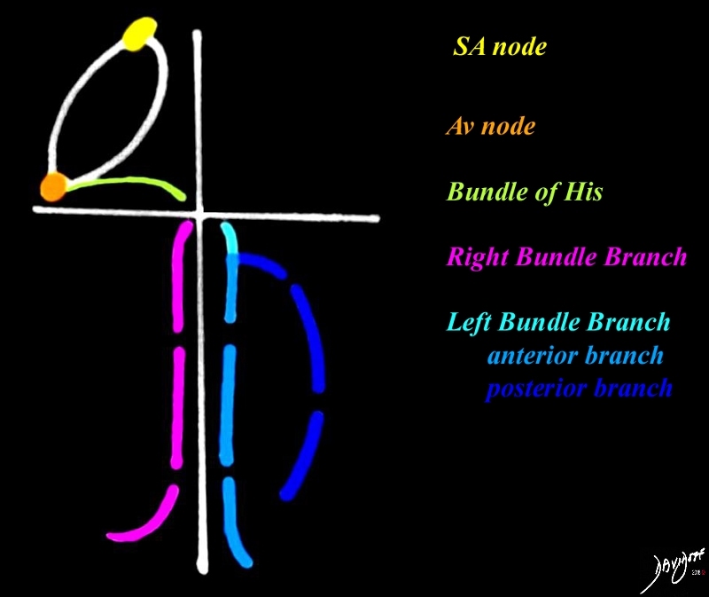
The SA node,(yellow) is positioned at the SVC RA junction, the A-V node at the IVC-RA junction, while the bundle of His (green) travels along the tricuspid annulus. At the level of the membranous septum it divides into the right bundle (pink) and left bundle(blue) and the left bundle divides into an anterior and posterior branch
Ashley Davidoff MD
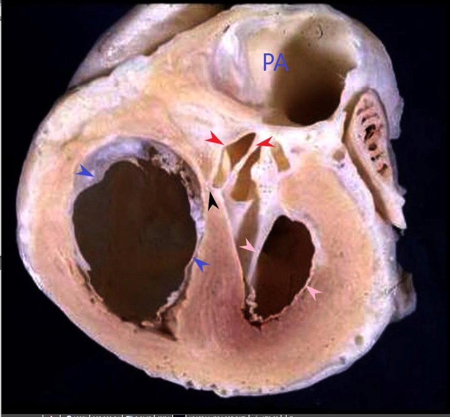
The anatomic specimen shows the right ventricle and the tricuspid valve (blue arrows), the left ventricle and the mitral valve (pink arrows) , with fibrous continuity with the aortic valve, and the region of the membranous septum (black arrow) at the crux of the heart
Ashley Davidoff MD
A Sense of the Cross in Axial Projection
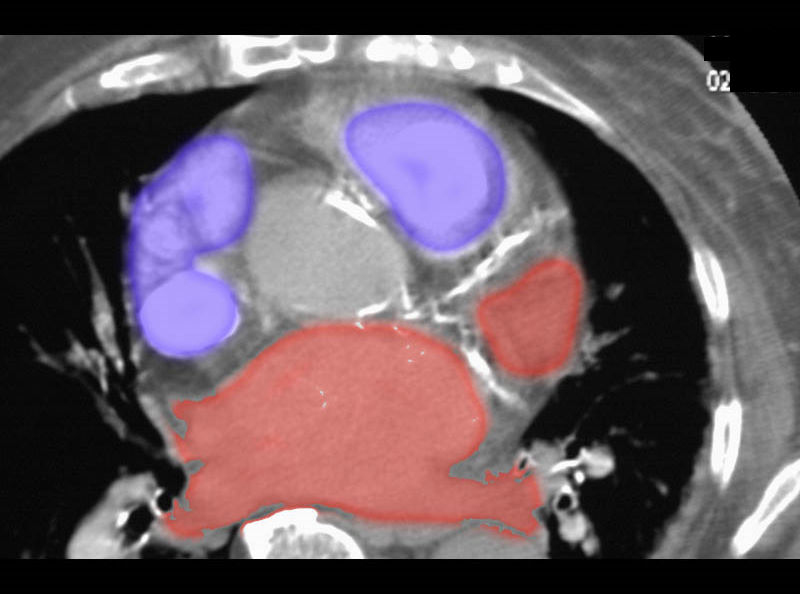
CT scan with coronary artery calcification provides a sense of the cross in a single plane in the axial projection. Note right sided structures are right and anterior
Ashley Davidoff MD
CORONARY ANATOMY
-
Coronaries Injected with Barium Anatomical Specimen
-
Coronaries Injected with Barium – Xray
-
Coronary Arteries in LAO on Angiography
-
GIF Practice
-
Coronaries on CT
Anomalous Origin
Other Congenital Anomalies
Things to Remember
LAD – septals 2/3
PDA 1/3
Dominance 70/20/10
SA nodal 55/45
Collaterals
Vieussens
Septals
Kugels
Inter -arterial Anomalous origins