- LAD aka widow maker
- Branch of the Left Main
- Size
- diameter 4-5mm
- length varies
- type-I – does not supply the left ventricular (LV) apex,
- type-2 – supplies part of the apex, the rest being supplied by the right coronary both,
- type3 – supplies the entire apex, and
- type-4 – supplies the apex and >25% of the inferior wall (wrap around).
- 50% circulation of the heart
- Shape
- Moustache
- Position
- Epicardial surface
- Sometimes a portion is intramuscular aka myocardial bridging
- relations
- Lymphatics of the heart travel with the Arteries
- Character
- Size
- Branches
- Septal branches supplying 2/3 of the septum
- Diagonals – supplying the anterior free wall of the LV
- Arc of vieussens Conal
- Anterolateral papillary muscle of RV
- Variations
- Bridging
- Absent Left MAin
- Coronary Fistulae: Coronary fistulae are rare in adults, but account for 50% of pediatric coronary anomalies. A fistula between a coronary artery and the cardiac cavity is called a coronary-chimeral fistula, that between a coronary artery and vein is called an arterio-venous fistula. Most fistulae are congenital. Acquired ones are usually iatrogenic or traumatic.
- Anomalous Origin: Anomalous origin of left coronary artery from the pulmonary artery is a rare malformation (incidence of 0.25–0.50%) in children with abnormal cardiac development leading to a mortality rate of 90% in unoperated infants.[4]
-
Left anterior descending: (coronary artery of Vieussens) The LAD courses in the anterior interventricular groove towards the apex of the heart . a direct continuation of the LM stem from the bifurcation nourishes the anterior and lateral wall of the left ventricle by coursing anteriorly and inferiorly towards the apex.
Course : The LAD a direct continuation of the LM from the bifurcation, adopts a S shaped course by passing to the left of the pulmonary trunk and then running anteriorly and inferiorly in the anterior interventricular groove towards the apex which it wraps around in as many as 80% of the cases to reach the inferior wall. It occasionally continues through the posterior interventricular groove, where it is known as Mouchet’s posterior recurrent interventricular artery. The left anterior descending artery (LAD) has the most constant origin, course and distribution and is usually divided into proximal, middle and distal segments.
Branches of LAD: The LAD gives rise to two main groups of branches. (1) The septal branches, which supply the anterior two-thirds of the septum, and (2) The diagonal branches, which lie on the lateral aspect of the left ventricle. A third set of branches are the right free wall branches.
-
DISTRIBUTION OF THE CORONARY ARTERIES
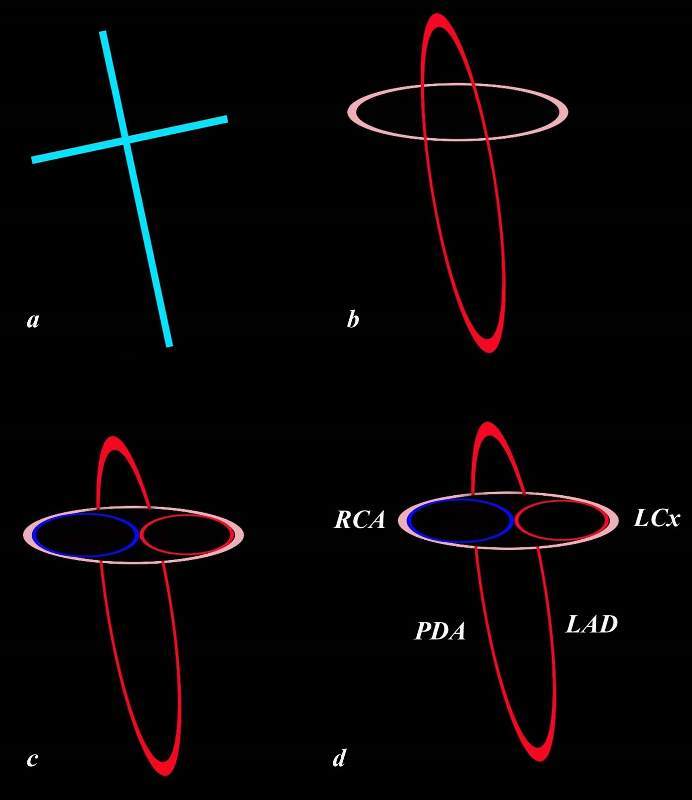
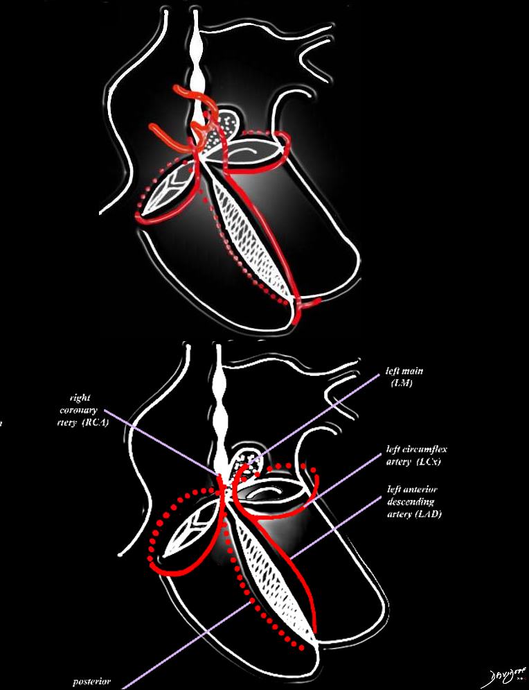
The heart has a left and right arterial system. The vessels are formed around the cross of the heart, “vertically” along the interventricular septum and interatrial septum, and “horizontally” in the atrioventricular (A-V) grooves. The left coronary artery arises from the left coronary ostium and supplies one branch, the left anterior descending artery, that travels anteriorly on the anterior aspect of the interventricular septum, and one branch that travels in the atrioventricular groove (left circumflex). The right coronary artery has a branch that courses along the right atrioventricular groove and usually continues as the posterior descending artery on the posterior aspect of the interventricular groove, and commonly gives rise to the A-V nodal branch, at the back of the crux of the heart, which travels in the vertical axis in the interatrial septum.
Courtesy of: Ashley Davidoff, M.D.
Post Mortem Barium Angiogram
Ashley Davidoff MD
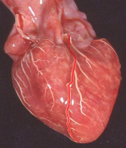
This is an anterior view of the heart showing the long LAD travelling within the expected course of the interventricular septum.
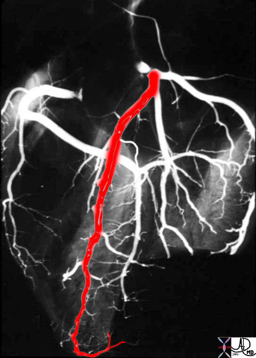
This post mortem angiogram in the LAO projection shows normal LAD overlaid in red with the PDA as the main vessels slightly to the hearts lt side. The septal branches from the PDA are well seen and the interventricular septum can be seen as a faint blush.
key words
cardiac heart coronary artery LAD diagonal conal infundibulum RVOT anatomy normal
Ashley Davidoff MD
33805aladP
44198d14 32134
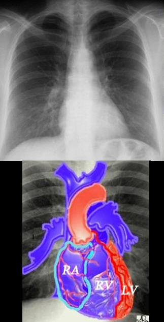
If we were to “crack open” the chest of the chest X-ray, the structures that would dominate this bloody, black and white scene, would be the right sided chambers. The right ventricle (RV) would be the dominant anterior chamber, and would form the dominant interface with the diaphragm. The right atrium (RA) would form the border with the right lung. The RA would of course be slightly posterior to the RV. The left border would be formed by the left ventricle. Most the left ventricle is hidden posteriorly in this view. The left anterior descending artery would be visible from this anterior view. It marks the position of the interventricular septum.
Ashley Davidoff MD
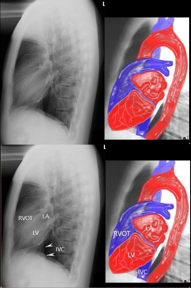
Ashley Davidoff MD

Normal Angiogram
Ashley Davidoff MD

Normal Angiogram
Ashley Davidoff MD
References and Links
Ibraheem Rehman; Afzal Rehman. Anatomy, Thorax, Heart Left Anterior Descending (LAD) Artery Stat Pearls

