- A Subset of Partial Voluming Artifact
- caused by
- small, high density structures such as
- artery calcifications and
- metallic objects,
- small, high density structures such as
- which appear larger than their true size
- due to
- limited spatial resolution,
- associated with the design tradeoffs between
- image noise and resolution, and with the
- partial volume averaging of different densities within a single voxel.
- associated with the design tradeoffs between
- with some motion artifact also contributing
- limited spatial resolution,
- leads to
- overestimation of luminal stenosis and
- result in inappropriate management.
- may consider not performing a CTA if the calcium score is greater than 100

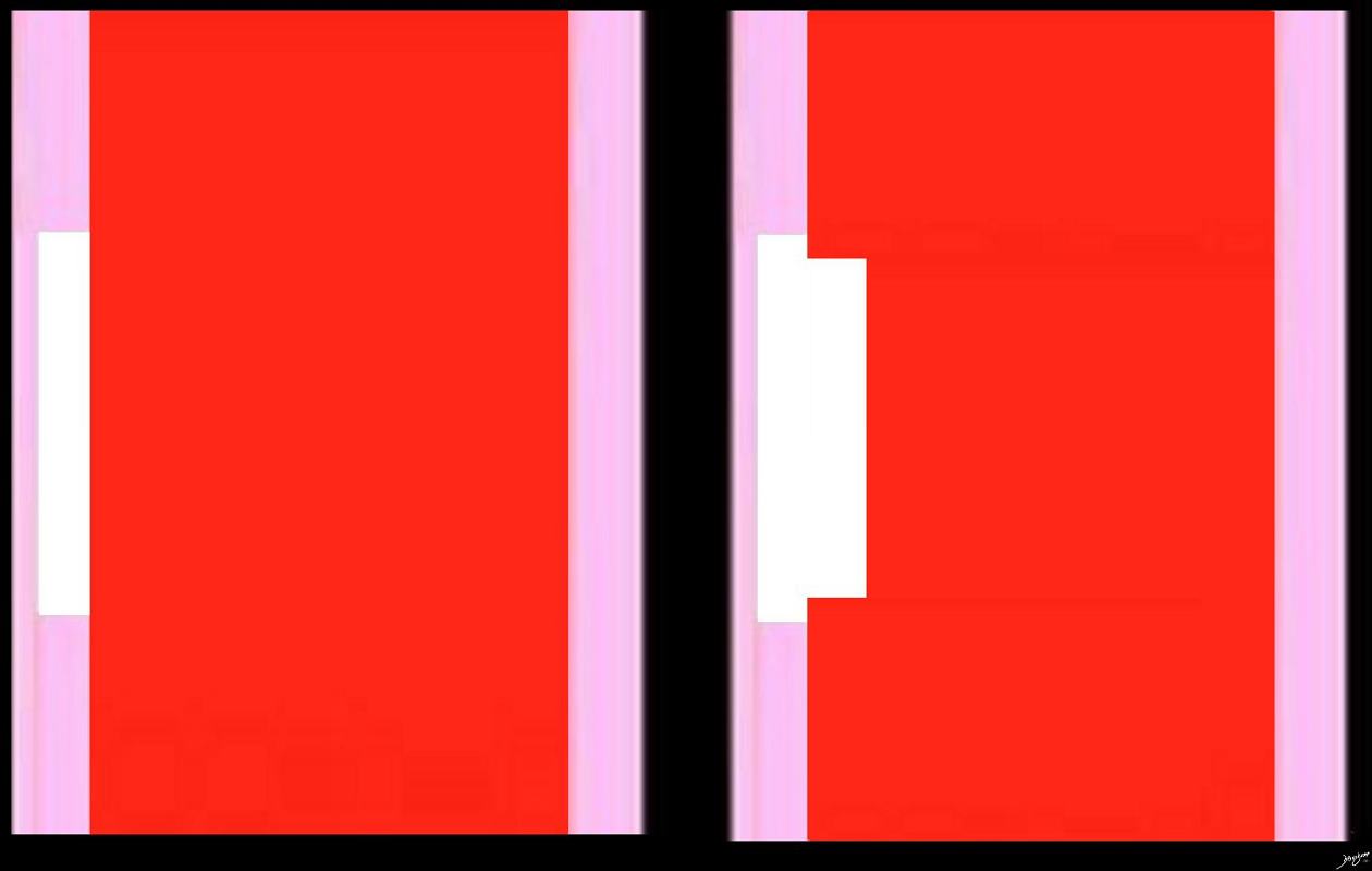
The small calcified plaque in the left image appears larger than it actually is in the right resulting in apparent impingement on the lumen when in actuality there is not. This is caused by blooming artifact because the pixel or voxels share structures of different density and the net density of a pixel is an average of the densities
Ashley Davidoff MD thecommonvein.net

The small calcified plaque in the top left image is an artistic version of the reality – calcified plaque that does not impinge on the lumen. The top right image is a CT in the ideal world recreated within the grid of pixels (voxels in 3D) The calcified plaque (white arrow) which shares a voxel space with the blood in the lumen) remains distinct and true to its size, demonstrating true lack of impingement on the lumen
The bottom row in gray scale shows what happens in a CT scan. The bottom left image shows what the CT would look like ie impingement by the plaque on the lumen. The bottom right explains the blooming artifact. The calcium of very high density (about 500HU) shares the voxel space with the blood which is soft tissue density (about 50 HU) and so the net density is about 450 HU. The voxel does not therefore reflect a true density and the lesion therefore appears to impinge on the lumen when, in actuality it does not. This is called the blooming artifact
Ashley Davidoff MD thecommonvein.net

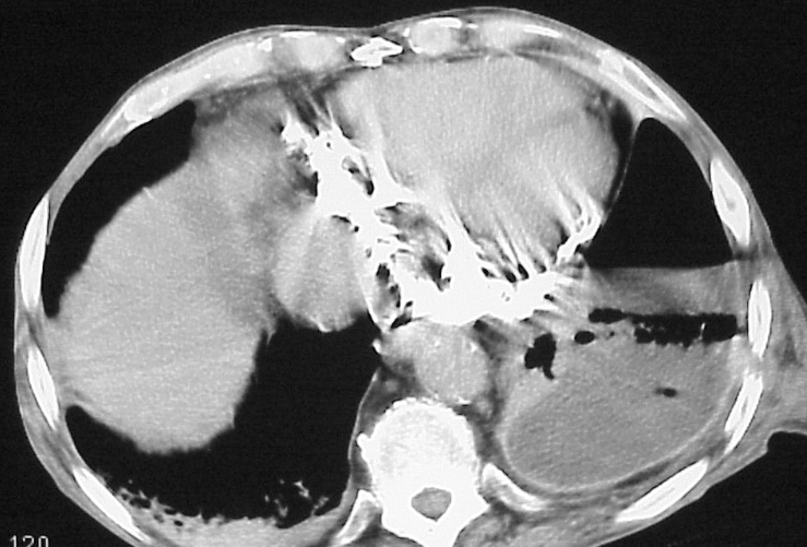
This CT scan of the heart shows unusually prominent blooming and mis-registration artifacts created by the calcification and the movement of the heart
key words
heart pericardium fx calcification serous pericardium fibrous pericardium dx calcific pericarditis probable viral in origin imaging
Ashley Davidoff MD
thecommonvein.net
Deblooming Algorithms

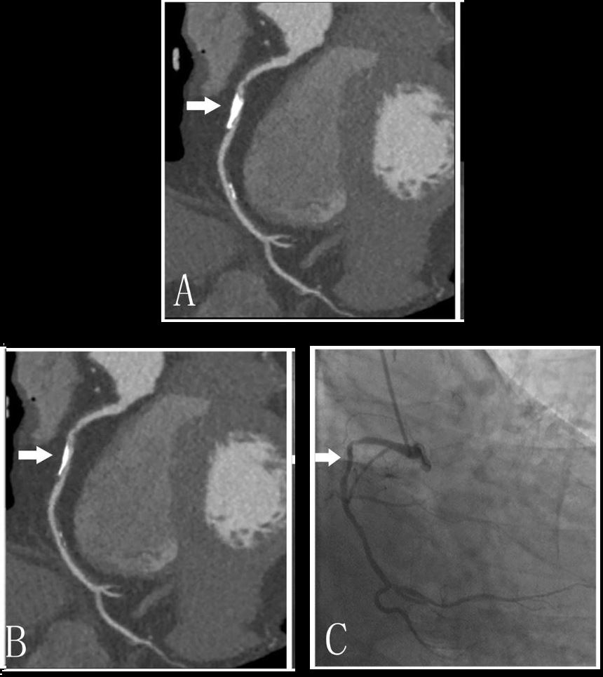
Ping Li, P et al Blooming Artifact Reduction in Coronary Artery Calcification by A New De-blooming Algorithm: Initial Study Scientific Reports volume 8, Article number: 6945 (2018)
