LGE MID MYOCARDIAL INFERO-LATERAL
MYOCARDITIS

Ashley Davidoff MD

Ashley Davidoff MD
Fabry Disease

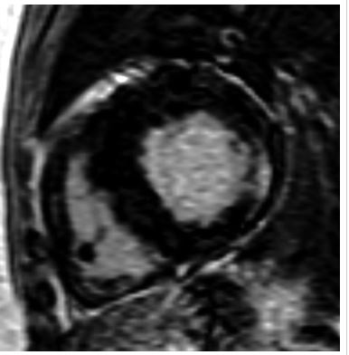
“Delayed enhanced images of 64-year-old heterozygotic woman with Fabry’s disease–related cardiac hypertrophy. Minimal thinning of basal segment of left ventricular inferolateral wall is associated with thick mesocardial striae of delayed enhancement shown on short-axis delayed enhanced images.”
De Cobelli et al Delayed-Enhanced Cardiac MRI for Differentiation of Fabry’s Disease from Symmetric Hypertrophic Cardiomyopathy
AJR Volume 192, Issue 3 2009
 LGE in FABRY DISEASE
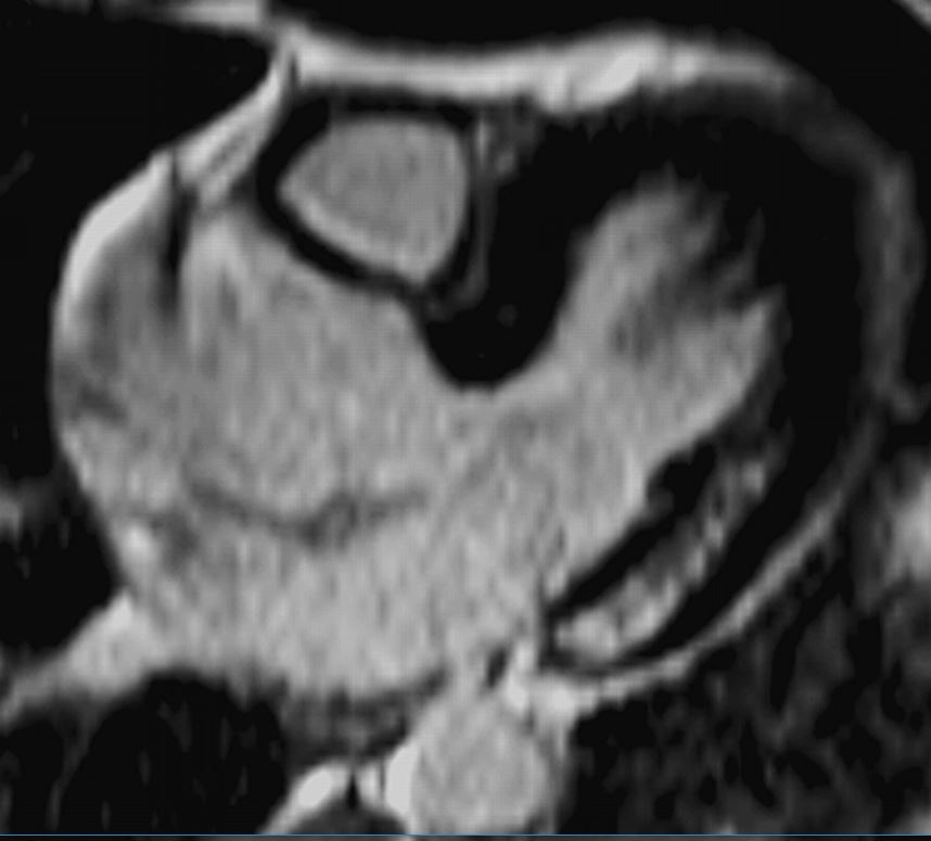
“40-year-old man with Fabry’s disease–related hypertrophy. Long-axis delayed enhanced images show typical pattern of delayed enhancement: thick striae involving inferolateral wall of basal segment of left ventricle in mesocardial distribution. Note sparing of subendocardial layer.
De Cobelli et al Delayed-Enhanced Cardiac MRI for Differentiation of Fabry’s Disease from Symmetric Hypertrophic Cardiomyopathy”
AJR Volume 192, Issue 3 2009
Nodular
Nodular
Sarcoidosis – Nodular Form

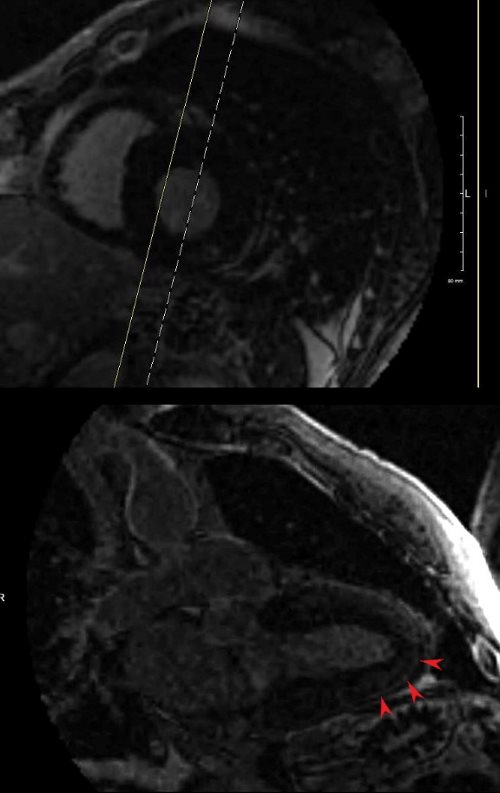
53 year old male with a clinical history of sarcoidosis and right bundle branch block. Echocardiography showed left ventricular hypertrophy and normal ejection fraction
MRI shows multifocal regions of LGE in the mid myocardium involving the inferobasal portion and inferolateral portion of the of the LV

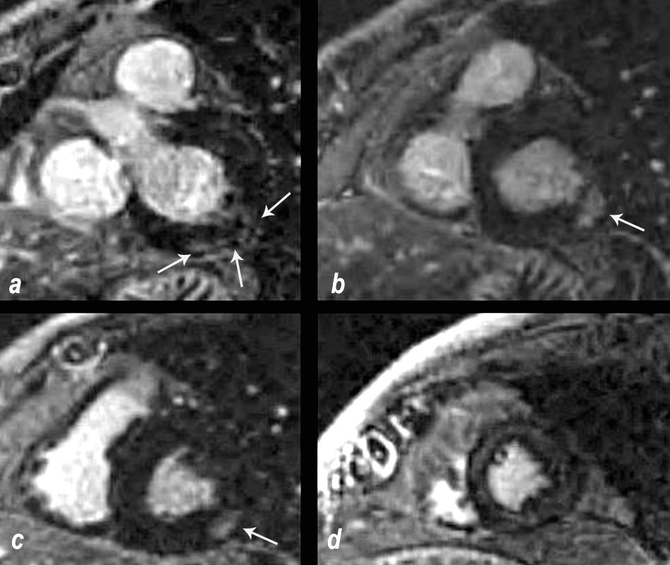
69 year old male presented with history of cardiomyopathy and atrial fibrillation
The findings on MRI are highly suggestive of sarcoidosis. There are multicentric foci of LGE in linear and nodular form in the mid myocardial and subepicardial layers and likely in the pericardium and myocardium of the right ventricle.
In this series images on the right (b,,d,,f) are magnified views of images on the left (a,c,e)
There are multicentric foci of LGE nodular form in the mid myocardial region (white arrowheads) linear LGE in the subepicardial layers (red arrowheads, b and f) in the pericardium (teal blue arrowhead) and myocardium of the right ventricle (yellow arrowhead e).
There was associated global hypokinesis of the LV with an EF of 40%, and increase in the LV mass of 120gms/ sq m
Ashley Davidoff MD
Fine Line Curvilinear
