
Goldfarb et al “We also found that fat deposition was predominately mid myocardial or mid epicardial, whereas results of prior autopsy and CT studies suggest that fat deposition almost always affects the subendocardium (28). MR imaging is unique for the study of fat deposition owing to its sensitivity to the off-resonance properties of fat, allowing fat-water separation imaging and the viewing of MI in all stages by using LGE imaging. ”
- POST MI FAT
Lipomatous Metaplasia
- Post MI,
- myofibroblasts produce collagen as a repair mechanism
- Sometimes myofibroblasts
- differentiate into adipocytes,
- which is laid down in the scar
- Consequence of myocardial infarction.
- At autopsy
- Lipomatous Metaplasia
- was seen in about 70% of scars associated with
- chronic ischemic heart disease
- Lipomatous Metaplasia is also
- a significant predictor
- heart failure,
- all-cause mortality,
- sustained ventricular arrhythmia
- Post MI,
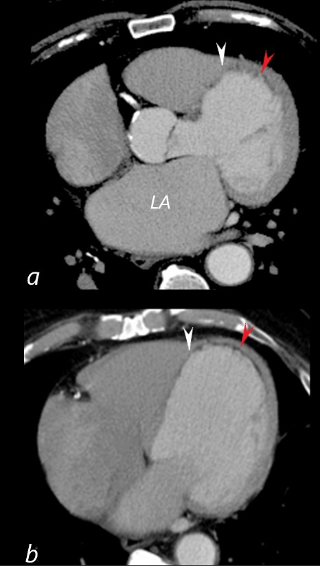
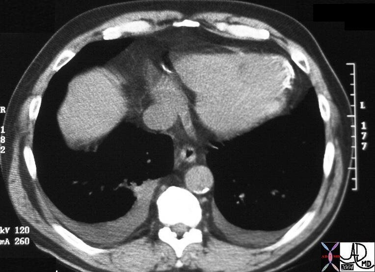
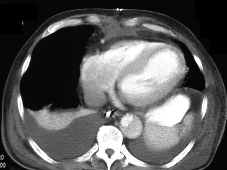
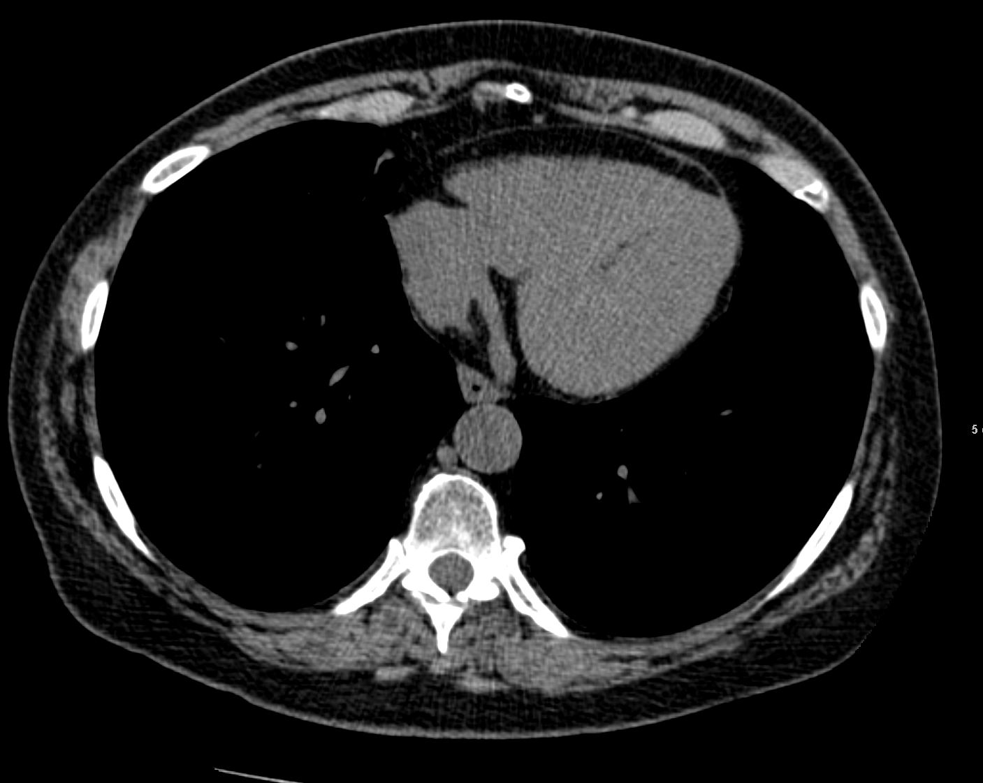
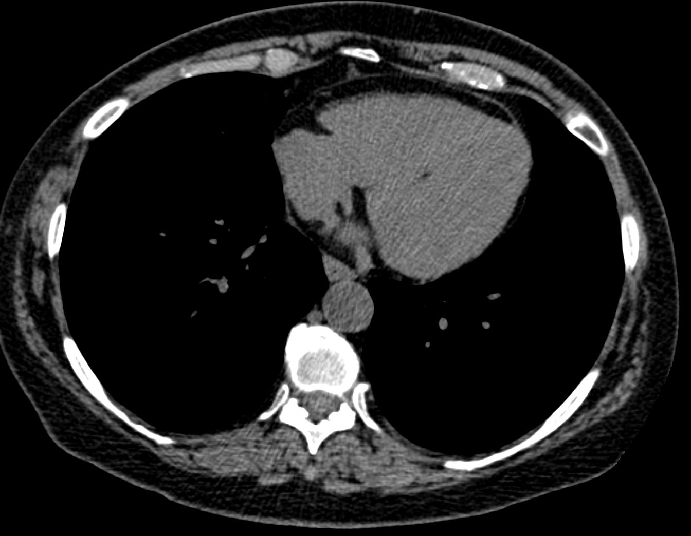
41 year old male presents with chest pain.
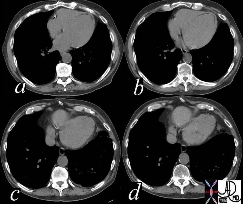
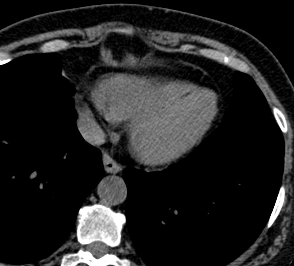
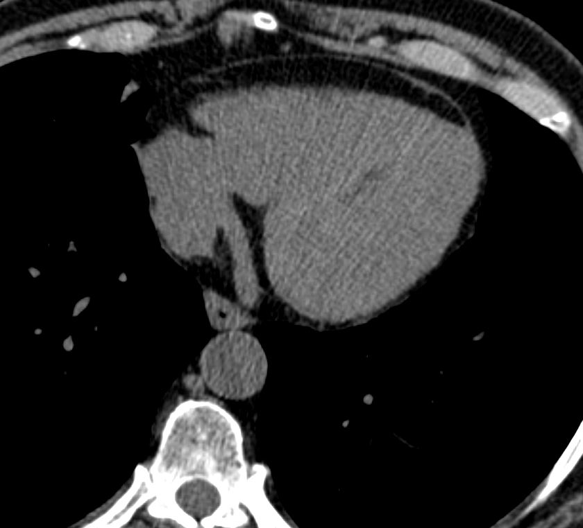
Axial CT with contrast at the level of the enlarged left atrium (LA) (a) reveals a dilated LV, with subendocardial fatty infiltration of the LV myocardium in the septum (white arrowhead), and apex red arrowhead). There is thinning of the myocardium in the septum and apex.
Image B is more inferior and taken at the level of the dilated RA and normal sized RV showing similar subendocardial fatty infiltration of the LV myocardium in the septum (white arrowhead), and apex red arrowhead). There is thinning of the myocardium in the septum and apex. The transverse dimension of the LV was 7.2 cms which is significantly dilated
These findings are consistent with myocardial infarction with and ischemic cardiomyopathy.
Ashley Davidoff MD
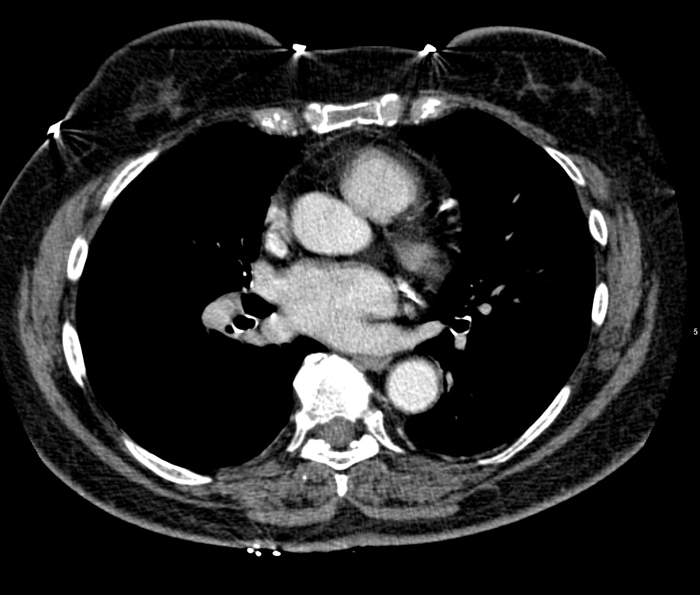
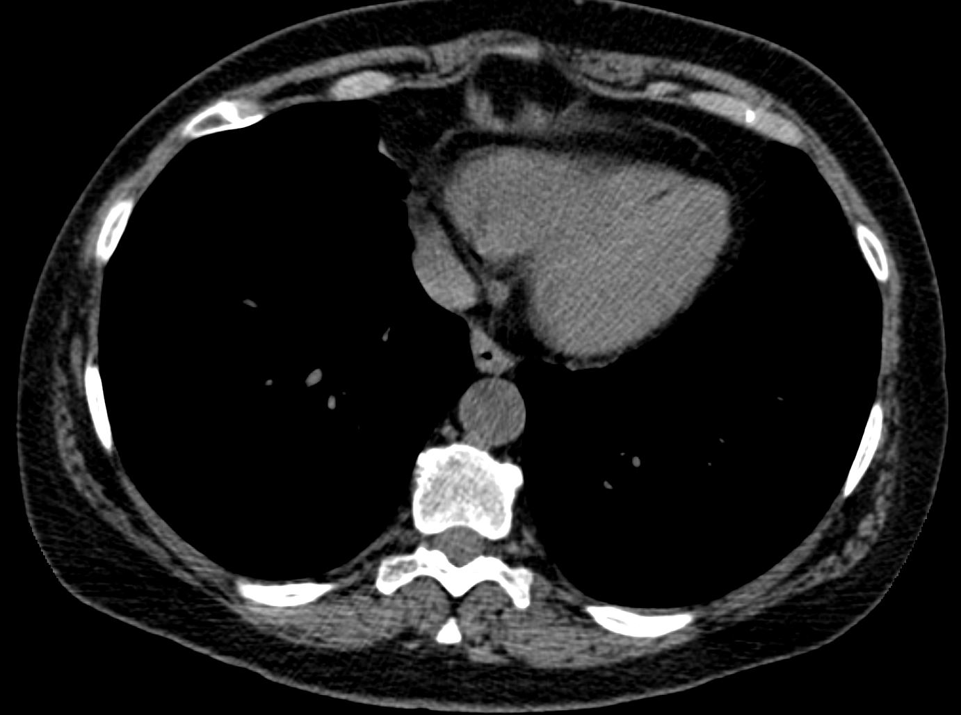
86943c03L has LCA aneurysm

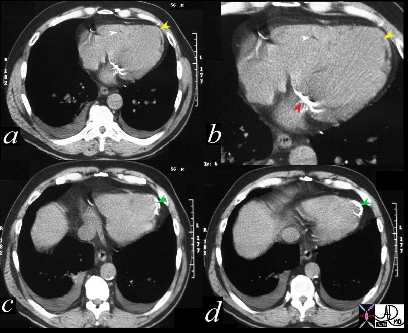
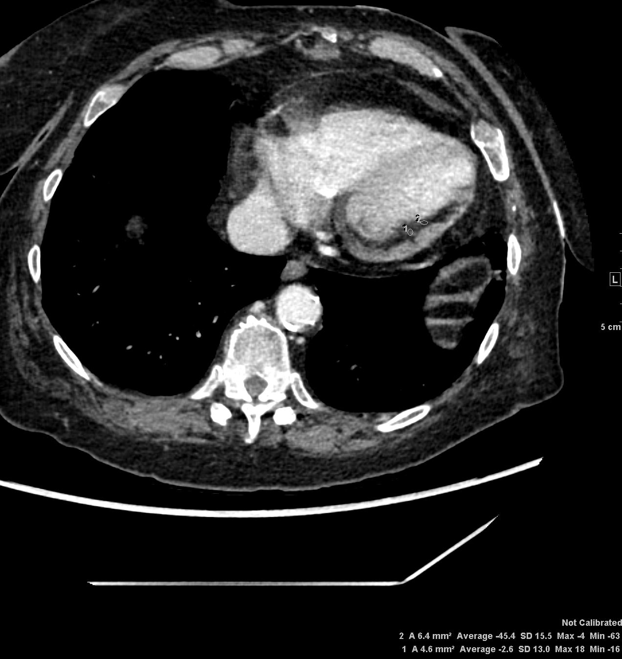
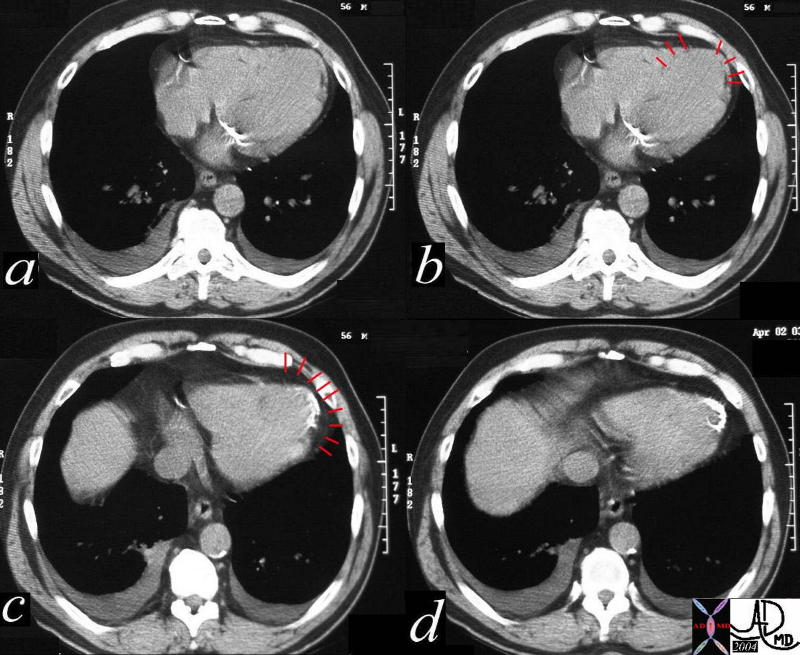
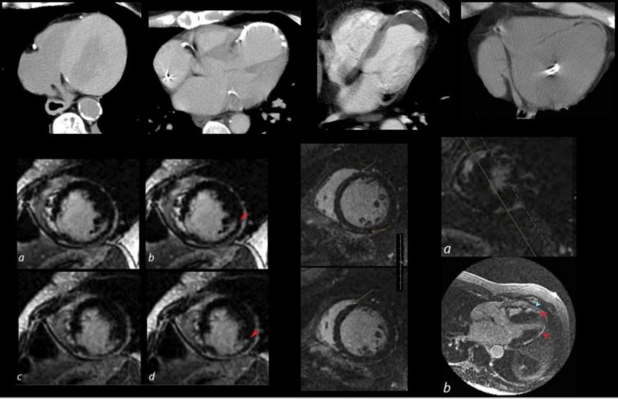
56 year old male with history of coronary artery disease. Axial CT through the heart shows apical curvilinear fat (yellow arrowheads, ( a and b) associated with apical myocardial dystrophic calcification (green arrowheads c and d) both indicating prior apical MI. In addition there mitral annular calcification (red arrowhead, b) and multifocal fatty deposits in the RV (white arrowheads, a and b) usually depicting age related degenerative changes,
Ashley Davidoff MD
The CommonVein.net
The CommonVein.net
The CommonVein.net
Ashley Davidoff
THECOMMONVEIN.net
Note also a dissection in the descending aorta
Ashley Davidoff
THECOMMONVEIN.net

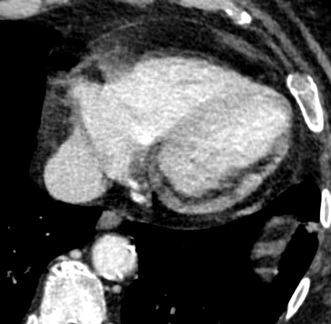
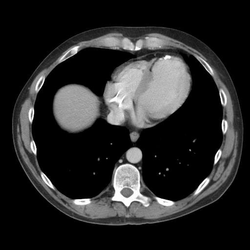
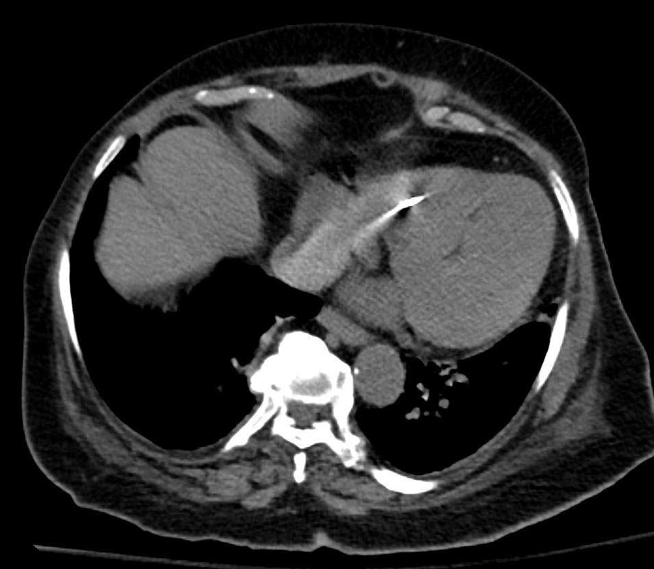
Dilated LV
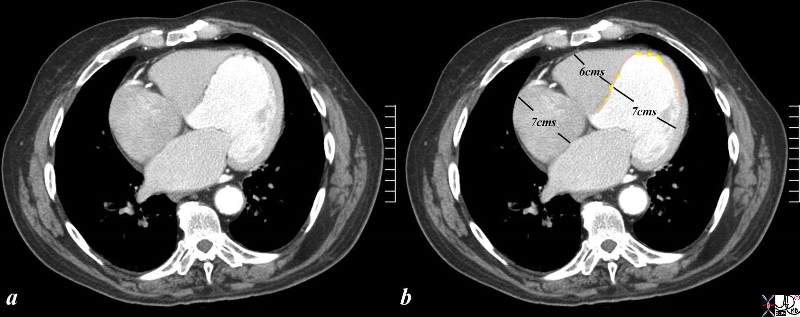
The CTscan is from a 76 year old man in whom the dominant finding is of left ventricular enlargement. The RA and RV are also enlarged based on this image, and LA was enlarged as well suggesting global cardiomegaly consistent with a cardiomyopathy. The clue to the cause of the enlargement is the segmental nature of the disease characterized by the asymmetry of the free wall and septum and the presence of fat (yellow overlay) in the thinned and probably scarred myocardium making ischemic cardiomyopathy the diagnosis. At the apex, there is there is a subtle calcification as well.
Courtesy Ashley Davidoff Copyright 2009 All rights reserved 86984c05.8s01
The collage reveals the variation in the tissue characterization of the LV in disease
Top row (from l to r) Patient with anemia showing low density of the blood and soft tissue density of the thickened myocardium. The 2nd image shows a calcified apical aneurysm. The 3rd image is a calcified aneurysm of the apex with thrombus in the lumen. The 4th image shows fat in the septum and the apex reflecting a previous MI in this region
2nd row 1st image is an MRI showing subendocardial delayed gadolinium enhancement (LGE) in a patient with a prior infarct of the inferolateral portion of the LV. The middle image shows a dilated LV with mid myocardial LGE in a patient with congestive cardiomyopathy. The last image shows multicentric linear and nodular LGE prominent in the subepicardial region and consistent with sarcoidosis.
heart 0086
Ashley Davidoff MD
Links and References
Goldfarb et al Myocardial Fat Deposition after Left Ventricular Myocardial Infarction: Assessment by Using MR Water-Fat Separation Imaging RadiologyVol. 253, No. 1