
Normal LVOT
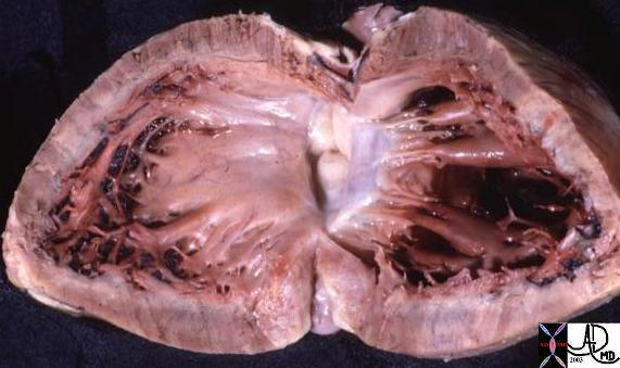
This anatomic specimen shows theleft ventricle ( LV ) with septal side to the patients right and free wall side to the patients left. Note the relatively smooth walled septal surface and the papillary muscle apparatus attached to the free wall side. There are two groups of papillary muscles neither of which are attached to the septum. The anterior leaflet of themitral valve (MV) is in full view and note that it is in fibrous continuity with the left and non coronary cusps of the aortic valve.
Courtesy Ashley Davidoff MD 01817