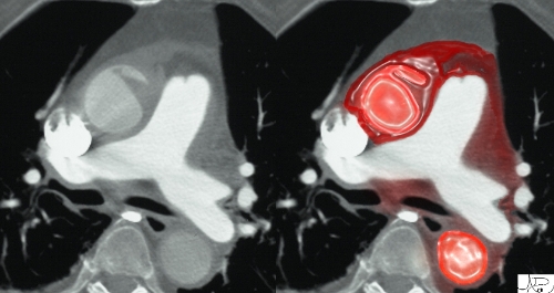
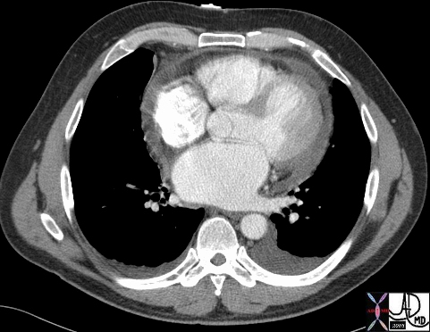
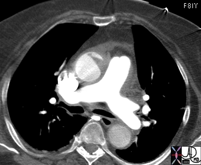
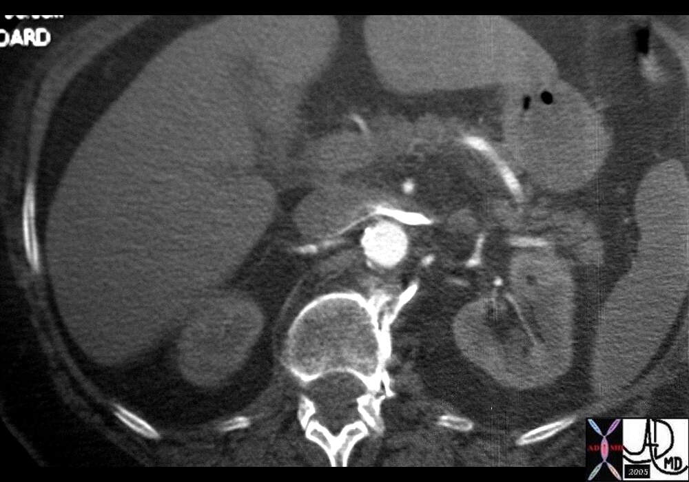
Pericardial tamponade is an abnormal accumulation of fluid in the pericardial space which results in diminshed filling of the atria and subsequently the ventricles complicated by reduction in cardiac output.
Th rapidity with which fluid accumulates is a major factor in the development of tamponade. Since blood usually accumulates rapidly hemorrhage into the pericardium is a common cause of tampnade. Penetrating trauma and hemorrhagig malignant effusions are therefore the common causes of tamponade.
- Major criteria:
- -Pericardial effusion (with pericardial fluid attenuation value greater than water)
- Enlargement of superior vena cava with a diameter similar to or greater than that of the adjacent thoracic aort
- Enlargement of inferior vena cava with a diameter greater than twice that of adjacent abdominal aorta
- Periportal lymphedema
- Minor criteria:
- “Flattened heart sign” – flattening of anterior surface of heart with decreased anteroposterior diameter
- -Compression of coronary sinus
- -Angulation/bowing of interventricular septum
- -Reflex of contrast within the IVC and/or azygos vein
- -Enlargement of hepatic or renal veins
- Potential complications:
- cardiogenic shock,
- myocardial infarction,
- arrhythmia,
- heart failure,
- embolism,
- wall rupture
Remember: findings seen individually are not specific, but when seen together strongly suggest the diagnosis of cardiac tamponade
22343

22355
CLINICAL
Pulsus paradoxus
defined as an inspiratory systolic fall in arterial pressure of 10 mm Hg or more during normal breathing
Echo
right atrium and ventricle – chamber collapse
25 percent have left atrial collapse

43695
43695 51M s/p ablation therapy for atrial fibrillation presents 3 weeks later with SOB pericardium fx small complex pericardial effusion fx thickened complex dx constrictive pericarditis caused by hemopericardium imaging radiology CTscan Courtesy Ashley Davidoff MD


