Buzzwords
- Pancarditis
- Mitral involvement is common – Nodules
- pericarditis most common reported
- Myocardial – increase ischemic and non ischemic
Pericarditis
less than 10 percent
most frequently in patients with active rheumatoid disease and other extraarticular manifestations.
Myocarditis —
Myocarditis usually associated with active articular disease
granulomatous fibrotic (more common in RA 46% )
involvement of the endocardium can cause MR
involvement of the conduction system can cause AV block
interstitial, (more common in SLE)
and with other nonarticular manifestations [49].
Myocardial abnormalities, were frequent in RA patients without known cardiac disease associated with higher RA disease activity, Inflammation in the pathogenesis of myocardial involvement in RA is implied
Valvular Disease
Rheumatoid nodules causing regurgitation
CAD
Ischemic cardiomyopathy rheumatoid vasculitis
increased risk – steroids
Conduction defects
Rheumatoid nodules causing
Amyloidosis
long standing effects
Drug Induced
Chloroquin and quinolones
MRI
Buzz
-
- patchy,
- mid-wall pattern,
- inferolateral

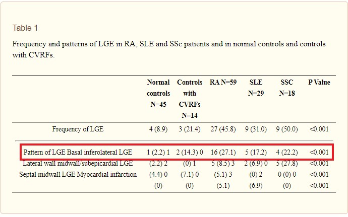
Ntusi et al Myocardial tissue characterisation with late gadolinium enhancement in rheumatoid arthritis, systemic lupus erythematosus and systemic sclerosis
J Cardiovasc Magn Reson. 2013; 15(Suppl 1): O47.

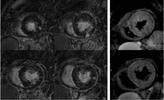
LGE noted as a linear pattern in mid myocardium on the inferolateral wall
Aisha T. Langford, Myocardial Inflammation Detected in Once Heart-Healthy RA Patients Cardiovascular Disease in Rheumatology, Rheumatoid Arthritis January 19, 2017

LGE noted as a linear pattern in mid myocardium on the anterior wall. linear
Aisha T. Langford, Myocardial Inflammation Detected in Once Heart-Healthy RA Patients Cardiovascular Disease in Rheumatology, Rheumatoid Arthritis January 19, 2017

LGE noted as nodular and linear changes in the mid myocardium and to some extent in the subendocardial and subepicardial regions extending the entire circumference
The second image shows diffuse T2 hyperintensity of the LV myocardium
Aisha T. Langford, Myocardial Inflammation Detected in Once Heart-Healthy RA Patients Cardiovascular Disease in Rheumatology, Rheumatoid Arthritis January 19, 2017

LGE noted as patchy nodular changes in the mid layer of the septum
Aisha T. Langford, Myocardial Inflammation Detected in Once Heart-Healthy RA Patients Cardiovascular Disease in Rheumatology, Rheumatoid Arthritis January 19, 2017

Ntusi et al Myocardial tissue characterisation with late gadolinium enhancement in rheumatoid arthritis, systemic lupus erythematosus and systemic sclerosis
J Cardiovasc Magn Reson. 2013; 15(Suppl 1): O47.
