Copyright 2019
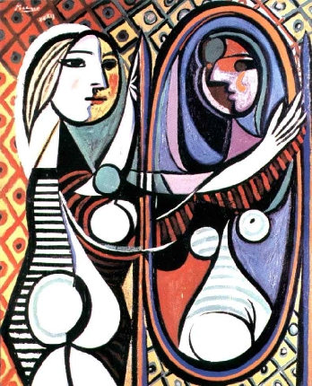
The structure that has enabled the two and a half billion beat pump has form that is equally marvelous and beautiful. Strictly it is made of four chambers and four valves, and each of the chambers is associated with a tubal system- either an inflow system of tubes or an outflow system. As one explores the structure it becomes fascinating to learn how each one of the components is uniquely designed and structured so that what appears at first to be a symmetrically designed system, turns out to be as asymmetrically symmetric as Picasso’s “Girl before a Mirror” (1932)
|
Girl Before the Mirror |
|
Despite the promised symmetry of “Girl before a Mirror,” 1932 we are surprised and fascinated by the asymmetric symmetry. The heart holds this same beauty. ©2001 Estate of Pablo Picasso/Artists Rights Society (ARS), New York |
In the scaffold model, we imagined the right atrium as a box on the top of the right ventricle, and the left atrium on top of the left ventricle. We have explored the heart as a whole up to now with some mention of the complexity of individual parts that make it up.
The Atria
As mentioned before, the right and left atria are structurally different beasts – as we say, “as different as “chalk and cheese.” Pectinate muscles, tenia saginata, and limbic bands all make the right atrium an extremely interesting chamber, as opposed to the more boring smooth-walled left atrium. Even as you look on the outside, the atrial appendages are different. The right appendage is more triangular in shape – like Snoopy’s nose – and the left appendage is shaped like a crooked index finger, or some say like the tip of the map of South America.
The left atrium (LA) is structurally the most boring of the four chambers. It does have a rather strange looking LA appendage , but its walls are mostly smooth. It has an interesting position in life in that it lies just beneath the crotch of the carina, and is almost central in the body with a mild bias to the left. Its posterior surface pats the distal esophagus, while its superior surface has the arm of the right pulmonary artery resting upon it.
The Ventricles
The right ventricle is oh-so-interesting as well, and so unlike the left ventricle. Firstly, as we have intimated, it is triangular in shape, relatively thin walled, but heavily trabeculated, and built to adapt to volume changes rather than pressure changes. The RV is really composed of two chambers – an inflow or sinus portion and an outflow or infundibular portion. Aristotle had suggested that the heart consisted of 5 chambers. Stella van Praagh, a wonderful teacher and friend, has translated the original Greek version of Aristotle’s work and firmly believes in her published work that Aristotle had the insight to recognize the right ventricular outflow chamber as the fifth chamber. The inflow and outflow components of the RV are anatomically and embryologically distinct. The RVOT is tubular in shape and elevates the pulmonary valve above aortic valve. The chamber is thin-walled, and twists around the aorta so that the pulmonary valve ends up to the left of the aortic valve. We are nearing the story of the “twist of the outflow tracts” Can you have the tricuspid valve in the left ventricle? The answer – no! What defines the name of the valve is the ventricle in which it lies. Hence the valve of the RV is the tricuspid, and the valve of the LV is the mitral (MV). The tricuspid valve is so named because it usually has 3 cusps, but this in fact is variable, and whether it has 3, 4, or 5, it is still named the tricuspid valve. The usual cusps are the anterior, septal and posterior cusps. The anterolateral papillary muscle of valve almost always originates from the moderator band which connects the free wall of the right ventricle with the septal wall. This papillary muscle can sometimes be used as an identifying feature of RV.
The left ventricle is a hollow muscular chamber of the heart. It has a conical shape and is the largest of the four chambers. The left ventricle lies in the middle mediastinum and is enclosed in the pericardium. It sits obliquely in the chest, behind the body of the sternum and the left inferior costochondral junctions, and projects from the midline into the left half of the thoracic cavity. It is responsible for taking oxygenated blood from the left ventricle via the mitral valve and propelling it forward through the aortic valve into the aorta to supply oxygenated blood to the tissues of the body. It consists of muscular fibers with unique shapes and unique function and also contains fibrous rings of the A-VB valves. It is covered by the visceral layer of the serous pericardium (epicardium) and lined by the endocardium. Between these two membranes is the muscular wall or myocardium. The fibrous rings surround the mitral and aortic orifices and serve for the attachment of the muscular fibers. The mitral ring is closely connected, by its right margin, with the aortic valve ring. The fibrous ring surrounding the orifices of the aorta serve for the attachment of the aorta itself and aortic valve.
-
Links and References
-
TCV

